Glomus Cancer
- Article Author:
- Oranus Mohammadi
- Article Editor:
- Manuel Suarez
- Updated:
- 4/26/2020 4:25:38 PM
- For CME on this topic:
- Glomus Cancer CME
- PubMed Link:
- Glomus Cancer
Introduction
Glomangiomas, or glomuvenous malformations (GVM), are rare cutaneous venous malformations that show glomus cells (undifferentiated smooth muscle cells, which are thermoregulatory units), along with venous system in histology.[1] Glomus cells specialized smooth muscle cells that regulate the temperature in the body.[2] Masson first described glomangiomas and Papoff further extensively studied.[3] There are three types of glomus tumors, classified based on its dominant component:
1. Solid: mainly glomus cells.
2. Glomangioma: mainly blood vessels.
3. Glomangiomyoma: mainly smooth muscle cells.[4] Glomangimyomas are further divided into (a) regional, (b) disseminated, and (c) congenital plaque-like.[5]
Glomangiomas usually present in multiples, often at birth or during childhood, and they do not involve the subungual region. A majority of glomangiomas are benign, although malignant cases have also been reported.[6][7] Rarely seen, the disseminated type distributes throughout the body.
Etiology
1. Inherited or familial (38% to 68% of glomangiomas):
Generally, it is autosome dominant with incomplete penetrance and variable expression, located in a 4--6--cm region of chromosome 1p21-22. Glomulin is the mutated gene which is located on the YAC and PAC maps. This gene includes 14 mutations in patients with this medical condition, which results in a loss of function. This loss of function increases cyclin E and c-Myc levels. In 60% of cases, one other family member is also affected. It can present at birth or later during adolescence.[8][7][9][10] 157_161del mutation is another documented mutation that may have a role in GVM malformations.[11]
Segmental type 2 is a variant of inherited glomangiomas. Initially they present with one primary lesion, followed by multiple distal lesions.
2. Sporadic or de novo mutation[12]:
It presents during birth.
Epidemiology
Glomangiomas are responsible for 1.6% to 2.0% of soft skin tumors and 20% of all glomus tumors. Plaque-like glomangiomas are very rare, with only 4 cases reported thus far. It is more predominant in the male gender. About one-third of the patients present before age 20.[8][13][14] 10% of cases are of the disseminated type.[10]
The most common reason for referral among vascular anomalies is venous malformations.[15]
Histopathology
History and Physical
It typically presents as purple skin lesions with a cobblestone pattern at birth. Lesions are usually bluish-purple, papular or nodular, hyperkeratotic, and 2 to 10 mm. The size and number of them are variable. These lesions are tender on palpation.[1][12] Pressure and cold trigger the pain. Areas rich with glomus bodies include the involved sites such as distal extremities, especially palms, wrists, forearms, feet, and subungual region. 75% of cases present in hands.[8] Visceral organ involvement is very rare, although it has been reported in the nasal cavity, mediastinum, gastrointestinal tract, respiratory tract, urogenital tract, and hepatobiliary system. Ventricular septal defects and transposition of the great vessels were reported in patients with GVM.[6][16][17] There is a case report of involvement of nerves with glomangioma, although normal human nerve is without glomus bodies.[18] Tracheal involvement is associated with dyspnea, hemoptysis, and retrosternal chest pain.[19]
Glomangiomas are larger and less well-circumscribed than solitary tumors. They have slow blood flow and usually grow over time.
The classic triad consists of the following : (a) hypersensitivity, (b) Intermittent pain, and (c) pinpoint pain.[20] Most of the time, glomangiomas do not present the classic triad.
The plaque-like type presents as indurated, nodular, or discolored lesions, which are non-tender and bleed with minor trauma. They are larger than the other types of glomangiomas.[14] This is the rarest type of glomangiomas.
Evaluation
The confirmation of the diagnosis is through histopathology.[8] If a tumor has atypical histology, immunohistochemistry assists in diagnosis. The role of smoothelin should be considered as it is an indicator of the smooth muscle cell.[21]
Electron microscopy shows glomus cells with dense bodies and smooth muscle myofibrils [1].
X-ray may show osseous defects.
MRI and color Doppler ultrasonography help define shape, size, and accurate location.[20][13] Dynamic time-resolved contrast-enhanced MR angiography can define the vascularity.[22]
Treatment / Management
The goal of treatment is to decrease the symptoms. It is difficult to treat the lesion when it reaches a specific size completely. For asymptomatic lesions, monitoring and observation are recommended. Surgery, electron-beam radiation, sclerotherapy with hypertonic saline or sodium tetradecyl sulfate, argon, flash lamp tunable dye laser (for multiple lesions), and CO2 lasers are different treatment modalities.[8][7] Excision therapy is the preferred treatment with painful lesions. Sclerotherapy was shown to be more effective in venous malformation than glomuvenous malformations. In cases of nasal involvement, endoscopic excision or surgery is recommended.[23][9][24] In cases of large glomangiomas that are difficult to excise, 1064-nm Nd:YAG laser is effective.[25] Also, positive results with Nd:YAG laser has been reported in symptomatic familial cases.[26][27]
External compression by elastic compressive garments worsens the pain.[12]
Differential Diagnosis
- Venous malformations: Glomangiomas are limited to the skin and mucosa. In contrast, other types of venous malformations can extend to deeper layers like muscles.[12]
- Schwannoma[28]
- Blue rubber bleb nevus syndrome (BRBNS)[7]: multiple visceral and cutaneous venous malformations, compressible lesions, sporadic, gastrointestinal bleeding is the reason for death
- Neuroma
- Hemangiopericytoma
- Angioleiomyoma
- Hamartoma
- Hemangioma[13]
- Subdermal mass
- Carcinoid tumors
- Hemangiopericytoma[19]
- Paraganglioma
- Maffucci syndrome: multiple subcutaneous vascular nodules on the toes and fingers
- Glomus tumor: (seen in the adult population), painful, more commonly involve nail beds, and genetic/histology is cellular dominant with glomus cell infiltration
- Spiradenoma
- Leiomyoma
- Venous malformation: compressible, painful
Prognosis
Complications
Recurrence after surgical excision is seen in 10% to 33% of cases.[9][13]
The chance of malignancy is very low. Risk factors for malignancy are the following: size greater than 2 cm, deep lesions, muscle, and bone invasion, and high mitotic activity.[14] Cases of metastasis were reported but are exceedingly rare.[10]
Nerve compression[29]
These lesions can be life-threatening due to the risk of growth, bleeding, or vital organ obstruction.[12]
One case reported Spitz nevi growing upon a congenital glomuvenous malformation. There is a theory that hyperemia in glomangioma can nourish the hair follicles. Mutated glomulin may also have a role in this case.[30]
Deterrence and Patient Education
It is recommended that patients return to their physician in case of further growth, bleeding, or recurrence.
Pearls and Other Issues
Sometimes it is challenging to diagnose GVM as there are different similar medical conditions, as discussed above.
Enhancing Healthcare Team Outcomes
Glomangiomas may exhibit signs and symptoms like papule or nodule.
It is important to consult with specialists, including dermatologists or surgery for the management of this medical condition. Specialty care nurses coordinate care and provide patient education. Radiologists may be involved during the assessment. Care can be enhanced by the interprofessional team. [Level 5]
(Click Image to Enlarge)
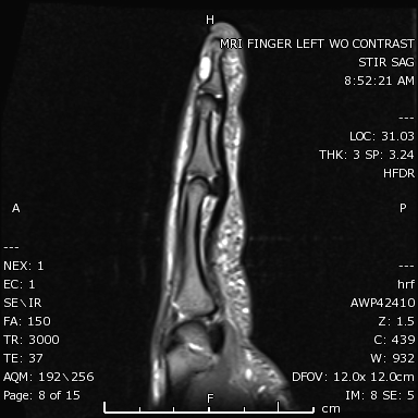
STIR sagittal image of the left index finger demonstrates a markedly hyperintense subungual mass.
Contributed by Hassana Barazi, MD.