Hallux Varus
- Article Author:
- Usama Munir
- Article Author:
- Ahmed Mabrouk
- Article Editor:
- Samer Morgan
- Updated:
- 8/26/2020 5:37:06 PM
- For CME on this topic:
- Hallux Varus CME
- PubMed Link:
- Hallux Varus
Introduction
Hallux varus is a clinical condition characterized by medial deviation of the great toe at metatarsophalangeal (MTP) joint. This condition may present with varying degrees of severity, causes, and symptoms. Adult acquired hallux varus deformity is commonly iatrogenic, commonly resulting from surgery for hallux valgus. (1,2,3)
A patient may have difficulty in walking and wearing shoes. Mild hallux varus can be managed with stretching exercises and splints. However, if the symptoms become significant and affect daily activities, then surgery should be considered.[1][2]
Etiology
Rarely, hallux varus is congenital. Flexible hallux varus may be found in newborns and reflects their intrauterine positioning. It corrects to valgus in early childhood when walking begins.[3]
More frequently this deformity develops after a surgical procedure for hallux valgus because of overcorrection, excessive lateral release, over-resection of medial eminence, over-plication of the medial capsule, zero-degree or negative intermetatarsal angle, or immobilization of the toe in excessive varus after surgery. Other causes include trauma and certain systemic inflammatory diseases such as psoriasis and rheumatoid arthritis.
- Congenital hallux varus is divided into primary and secondary pathologic deformity. Primary hallux varus is rare and related to an overactive abductor hallucis. Secondary hallux varus is related to great toe polydactyly, a delta phalanx longitudinal epiphyseal bracket syndrome, and metatarsus adductus.
- Medial insertion of the abductor tendon causes primary dynamic infantile hallux varus.
- Acquired adult hallux varus is an inflammatory arthropathy that includes psoriatic and rheumatoid arthritis. The mechanism of arthropathies combines destruction of the articular surfaces by distention of the joint capsule with subsequent laxity of the collateral ligaments, intrinsic muscular contracture, and pannus.
- Traumatic hallux varus occurs with sports injuries secondary to rupture of the lateral collateral ligament and conjoined tendon.
Spontaneous idiopathic hallux varus is noted incidentally, and the cause is not usually demonstrable.
Congenital hallux varus is typically due to connective tissue disorders (i.e., Marfan syndrome and Ehlers-Danlos syndrome) or is associated with Down syndrome and neuromuscular disorders (i.e., cerebral palsy).
Epidemiology
The incidence of iatrogenic postoperative hallux varus varies from 2% to 14% after corrective surgery for hallux valgus deformity. Crescentic osteotomies have an overall varus rate of 10%. However, the incidence of idiopathic, congenital/infantile, traumatic, and otherwise acquired hallux varus is unknown.[4]
Pathophysiology
With a chevron osteotomy, if the capital fragment is excessively displaced lateralward, a hallux varus deformity can develop. Likewise, with a proximal osteotomy, the distal segment can be translated too far laterally. For classical McBride procedure, the fibular sesamoid is excised which causes MTP joint hyperextension, interphalangeal (IP) joint flexion and medial deviation of the hallux.
With time, the deformity becomes fixed, and it is difficult for the patient to obtain comfortable footwear. The deformity usually manifests itself as the medial deviation of the great toe, supination of the phalanx and claw tow deformity.
Anatomically, cadaveric biomechanical studies reveal the restraints in descending order are the lateral capsule, the adductor hallucis, and the lateral flexor brevis tendon.
History and Physical
The hallux varus deformity if often asymptomatic. Some patients complain of deformity and have difficulty in wearing shoes, instability, decreased range of movement, and weakness with push-off. Pain indicates an underlying arthritic process. Patients may present with chronic pain, difficulty walking and standing for long, foot weakness, ingrowing toenails, limited MTP joint range of motion, swelling of the foot, and occasionally redness/ulceration of the big toe. The symptoms become worse when the patients wear closed-toe shoes that crowd the toes. The most common cause of pain in hallux varus is irritation of the deformed toe due to a poorly fitting shoe
A thorough assessment focusing on bone and joint deformity, joint flexibility and integrity, and soft-tissue balance. The following may be noted on physical examination:
- Bone deformity: Varus orientation of the great toe. Medial displacement of the medial sesamoid.
- Joint flexibility and Integrity: Dorsal contracture of the MTP joint with or without IP joint contracture. The degree of extension of the first MTP joint should be analyzed taking into consideration whether weight-bearing and the dynamics of ambulation increase the deformity or not.
- Soft tissue balance: Medial displacement of the extensor hallucis longus with a bowstring deformity. The plantar surface should be examined for any callosities.
Evaluation
Blood tests are only needed if one is expecting an infective or inflammatory process.
Weightbearing radiographs of both feet, including anteroposterior, lateral, and sesamoid, assess the degree of varus, the intermetatarsal and interphalangeal angles, absent lateral sesamoid, excessive medial eminence resection, the position of the sesamoids relative to the metatarsal head, and degenerative changes in the metatarsophalangeal or interphalangeal joints.
Treatment / Management
Non-operative treatment includes shoe stretching and modification. Shoes with wide toe boxes and padding over bony prominences should be recommended. For early postoperative varus deformities after hallux valgus correction surgery, taping or splinting the toes can be effective. This should be continued for 12 weeks until soft tissue healing. If there is persistent pain or inability to wear shoes, surgery is indicated.[5][6][7][8]
Aims of surgery include restoring and/or maintaining normal gait pattern and weight-bearing mechanism, realigning of sesamoids, correcting of deformity in the sagittal and transverse plane, and preserving the first MTP joint range of motion, if possible.
Operative treatment: Soft tissue or bony procedure.
The following should be considered to plan surgery:
- The type of deformity flexible or rigid.
- The degree of deformity.
- Presence of arthritis in the first metatarsophalangeal joint.
As a rule, the flexible deformity can be corrected with a soft tissue procedure. Lengthening of the medial capsular structures may be sufficient if the deformity is not too advanced. For advanced but flexible deformities the following procedures have been described either alone or in combination:
- Adductor hallucis tendon re-attachment with medial release
- Abductor hallucis tendon transfer on the base of the lateral base of the proximal phalanx in combination with reattachment or reefing of the conjoined tendon in the webspace or transfer of a part of EHB or EHL under the transverse intermetatarsal ligament to the distal metatarsal neck.
- Abductor hallucis tendon transfer to the lateral aspect of the proximal phalanx in combination with medial capsule release and medial sesamoid mobilization[9].
- Medial release without or with IP joint arthrodesis
- Split extensor hallucis brevis transfer and reverse Akin procedure.
- Tenodesis of the EHB tendon in combination with the release of the medial soft-tissues[10][11].
- Extensor hallucis longus (EHL) transfer to the proximal phalanx to act as a dynamic stabilizer [12]. This is done in combination with interphalangeal arthrodesis.
- Keller resection arthroplasty
- Implant arthroplasty
Contraindications for tendon transfer procedures:
- Arthropathies (Degenerative and inflammatory).
- Active infection.
- Neurovascular compromise (e.g peripheral neuropathy, peripheral vascular diseases).
- Excessive resection of the medial eminence.
- Fixed deformity of the metatarsophalangeal joint.
In cases with rigid deformity, deformity with limited first MTP joint motion, or presence of arthritic changes in the first MTP joint, arthrodesis of the first MTP joint is considered.
Lateral closing wedge osteotomy +/- repair of lateral ligaments can be offered in cases of iatrogenic hallux varus due to overcorrection of a hallux valgus deformity with either metatarsal or proximal phalangeal osteotomy.
Prognosis
Surgery improves the overall position of the hallux but not necessarily its motion. Salvage procedures may be necessary and corrective iatrogenic hallux varus procedures are 60% to 80% effective.
Complications
Potential complications of hallux varus surgery include the following:
- Soft tissue complications, including infection, impaired wound healing and nerve damage or irritation.
- MTP joint instability
- Recurrence of hallux varus
- Undercorrection or overcorrection
- Avascular necrosis of the metatarsal head
- Stiffness
- Hardware failure
- Progression of degenerative changes in the MTP joint
- Shortening of the medial column
- Transfer metatarsalgia
Enhancing Healthcare Team Outcomes
Hallux varus is a relatively common foot deformity seen in clinics. Because there is no good treatment, early diagnosis and changes in shoe wear are key. The condition is best managed by an interprofessional team that includes a podiatrist, orthopedic surgeon, physical therapist and nurse practitioner. Changing shoe wear is beneficial but many patients have a significant deformity that requires surgery.
Surgery improves the overall position of the hallux but not necessarily its motion. Salvage procedures may be necessary and corrective iatrogenic hallux varus procedures are 60% to 80% effective.[13][14]
(Click Image to Enlarge)
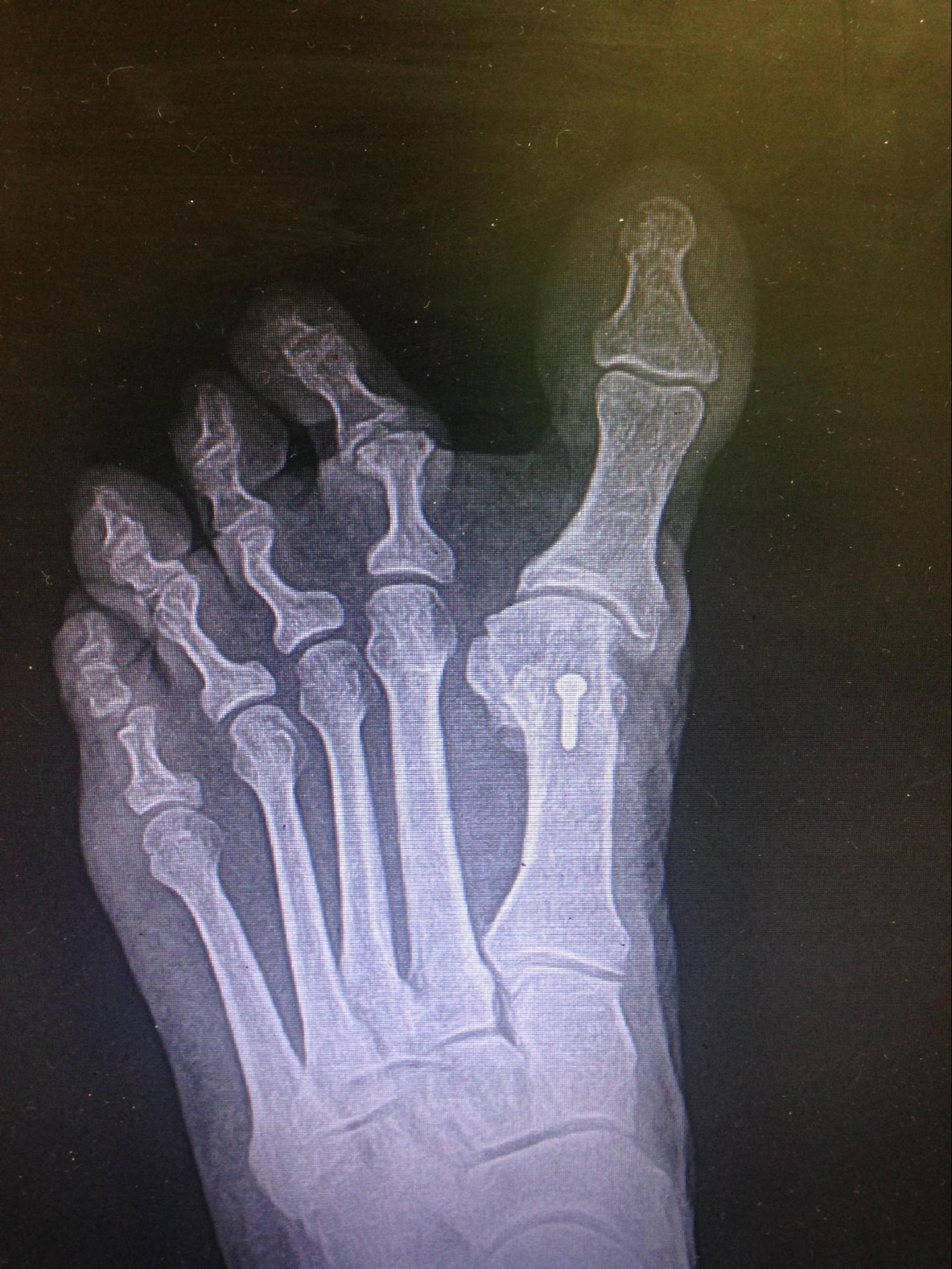
Hallux Varus Iatrogenic Hallux varus or abduction in the transverse plane secondary to surgical overcorrection of hallux valgus (bunion).
Contributed by Mark A. Dreyer, DPM, FACFAS