Hand Foot And Mouth Disease
- Article Author:
- Amanda Guerra
- Article Author:
- Emily Orille
- Article Editor:
- Muhammad Waseem
- Updated:
- 9/22/2020 12:13:42 AM
- For CME on this topic:
- Hand Foot And Mouth Disease CME
- PubMed Link:
- Hand Foot And Mouth Disease
Introduction
Hand, foot, and mouth disease (HFMD) is a common viral illness usually affecting infants and children but can affect adults. The infection usually involves the hands, feet, mouth, and sometimes, even the genitals and buttocks. The cause of hand, foot and mouth disease is coxsackievirus A type 16 in most cases, but the infection can also be caused by many other strains of coxsackievirus. In the western Pacific, hand, foot, and mouth disease has been linked to enterovirus. The coxsackievirus is a member of the Picornaviridae family which includes non-enveloped single-stranded RNA viruses.[1][2][3]
Etiology
Epidemiology
This viral infection is not indigenous to one area in particular but occurs worldwide. As children (particularly those younger than seven years of age) tend to be infected at a higher rate than adults you can see outbreaks in daycares, summer camps or within the family. These outbreaks are usually during the summer and early autumn.
Hand, foot, and mouth disease occurs at an equal frequency in both genders, but older epidemiological data seem to suggest that the frequency of infection is slightly higher in males.
The majority of people infected with coxsackievirus are children under the age of 10. Because the virus is shed in the stools for many weeks, some studies indicate that family members and close contacts are also at risk for developing hand, foot and mouth disease.
Pathophysiology
The spread of the human enterovirus is mediated by oral ingestion of the shed virus from the gastrointestinal or upper respiratory tract of infected hosts, or via vesicle fluid or oral secretions. After ingestion, the virus replicates in the lymphoid tissue of the lower intestine and the pharynx and spreads to the regional lymph nodes. This can be spread to multiple organs including the central nervous system, heart, liver, and skin.
History and Physical
Hand, foot and mouth disease can start with a low-grade fever, reduced appetite, and general malaise. The most common hand, foot, and mouth disease presenting symptom is usually mouth or throat pain secondary to the enanthem. The presence of vesicles surrounded by a thin halo of erythema, eventually rupturing and forming superficial ulcers with a grey-yellow base and erythematous rim. The exanthem can be macular, papular or vesicular. The lesions are non-pruritic, and typically not painful. The exanthem involves the dorsum of the hand, feet, buttocks, legs, and arms.
Hand, foot and mouth disease can also present with atypical features like concomitant aseptic meningitis. Enterovirus infections that cause hand, foot, and mouth disease are notorious for involving the central nervous system (CNS) and may cause encephalitis, polio-like syndrome, acute transverse myelitis, Guillain-Barre syndrome, benign intracranial hypertension and acute cerebellar ataxia.
Evaluation
The diagnosis of hand, foot, and mouth disease is usually made clinically. The virus can be detected in the stool for about 6 weeks after infection, however, shedding from oropharynx is usually less than 4 weeks. Light microscopy of biopsies or scrapings of vesicles will differentiate hand, foot, and mouth disease from varicella zoster virus and herpes simplex virus.[7][8]
While serology is not sensitive to make a diagnosis of hand, foot, and mouth disease, levels of IgG can be used to monitor recovery. In some centers, serology is used to differentiate enterovirus 71 from coxsackievirus as this has prognostic significance. Today, polymerase chain reaction assays are available in most centers to confirm the diagnosis of coxsackievirus.
Treatment / Management
Hand, foot, and mouth disease is a mild clinical syndrome and will resolve within 7 to 10 days. Treatment is primarily supportive. Pain and fever can be managed with NSAIDs and acetaminophen. Making sure the patient remains well hydrated is important. Additionally, a mixture of liquid ibuprofen and liquid diphenhydramine can be used to gargle with which helps coat the ulcers, ameliorating the pain.[9][10]
Ribavirin, quinacrine, and amantadine have all been used off-label to treat severe cases of enterovirus 71 induced hand, foot, and mouth disease disease.
Over the past decade, researchers have developed specific treatments to manage enterovirus 71 induced hand, foot, and mouth disease because of its severe neurological complications. So far, no drug has been approved, but promising novel agents include molecular decoys, translation inhibitors, receptor antagonists and replication inhibitors. One new antiviral agent that has shown promise in the treatment of enterovirus 71 is pleconaril.[11]
Differential Diagnosis
- Enteroviruses
- Erythema multiforme
- Herpangina
- Herpes simplex
- Herpes zoster
- Kawasaki disease
- Toxic epidermal necrolysis(TEN)
- Viral pharyngitis
Prognosis
The prognosis for the majority of patients with hand, foot, and mouth disease is excellent. Most patients recover within a few weeks without any residual sequelae. The acute illness usually lasts 10 to 14 days, and the infection rarely recurs or persists. However, some patients with hand, foot, and mouth disease may develop serious complications which include the following:
- Persistent stomatitis which is associated with painful ulcers. The pain can be severe enough to limit intake of food and dehydration can result, especially in young children.
- Aseptic meningitis can occur, but this is more common with enterovirus 71. This particular virus is associated with a higher rate of neurological involvement compared to coxsackievirus. The individual may develop acute cerebellar ataxia, polio-like syndrome, encephalitis, benign intracranial hypertension and Guillain-Barre syndrome. It is believed that the virus induces damage to the gray matter which then results in the motor dysfunction.
- Very rarely coxsackievirus can cause interstitial pneumonia, myocarditis, and pulmonary edema.
- Some studies indicate that coxsackievirus infections may also be associated with spontaneous abortions.
Pearls and Other Issues
The majority of patients with coxsackievirus-induced hand, foot, and mouth disease are treated as outpatients, but those who have CNS involvement may require admission for close monitoring. These patients often require imaging studies of the brain to guide treatment and recovery. Infants may develop dehydration, especially if they develop painful oral ulcers and may require Intravenous hydration. Admission is highly recommended in any infant with hand, foot, and mouth disease who shows signs of severe disease and lethargy. The virus is shed in the stools for a few weeks; hence, patients should be educated about hand washing and maintenance of good personal hygiene.
Enhancing Healthcare Team Outcomes
Cases of HFMD are on the rise and clinicians need to know how to make the diagnosis. Because many recent cases have involved the brain, an interprofessional team that includes a neurologist, internist and an infectious disease expert should be involved. Patients with signs of encephalitis need to be monitored by the nurses.
The outcomes for most patients with HFMD are excellent, with full recovery occurring within 7-21 days.
(Click Image to Enlarge)
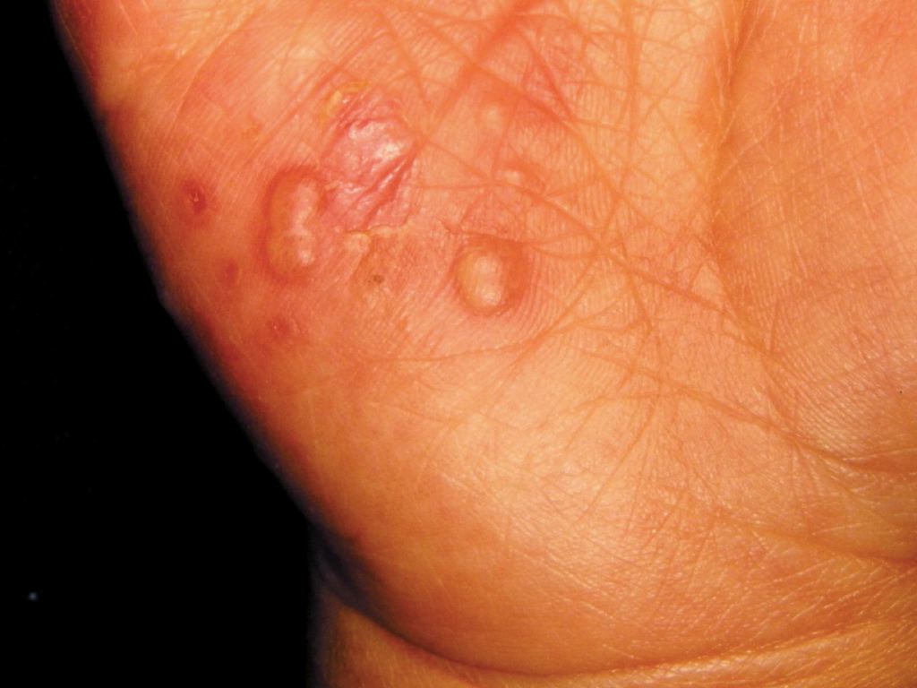
Hand and Mouth
Contributed by DermNetNZ