Horner Syndrome
- Article Author:
- Zalan Khan
- Article Editor:
- Pradeep Bollu
- Updated:
- 5/10/2020 6:48:55 PM
- For CME on this topic:
- Horner Syndrome CME
- PubMed Link:
- Horner Syndrome
Introduction
Horner syndrome is a rare condition classically presenting with partial ptosis (drooping or falling of upper eyelid), miosis (constricted pupil) and facial anhidrosis (loss of sweating) due to a disruption in the sympathetic nerve supply. It is primarily acquired following damage to the sympathetic nerve supply, but rare cases of congenital forms have been seen. Treatment is centered around identification and appropriate management of the underlying secondary cause.
The syndrome has several names such as Bernard-Horner syndrome (French-speaking countries), Horner syndrome (English speaking countries), oculosympathetic palsy and Von Passow syndrome (Horner syndrome in association with iris heterochromia).
The syndrome was first described by Francois Pourfour du Petit in 1727 when considering results from an animal experiment involving resection of intercostal nerves and subsequent changes seen in the ipsilateral eye and face. It was outlined more thoroughly by the French physiologist, Claude Bernard in 1852, followed by several physicians who offered different interpretations.
The condition was formally described and later named after a Swiss ophthalmologist Johann Friedrich Horner in 1869. [1][2][3][4]
Anatomy
Understanding the sympathetic innervation of the eye is vital to understanding the features of this syndrome. The nerve supply is constituted by three different neurons, starting from the posterolateral hypothalamus and ending as the long ciliary nerves to supply the iris dilator and superior tarsal muscles (Muller muscle).
The first-order neurons originate from the hypothalamus and descend through the midbrain and pons uncrossed, terminating at the C8-T2 level of the spinal cord in the intermediolateral cell columns (ciliospinal center of Budge). Second-order preganglionic neurons exit at the T1 level of the spinal cord to enter the cervical sympathetic chain where the fibers ascend to synapse in the superior cervical ganglion at C3-C4 level.
Third-order, postganglionic fibers, branch off into the sudomotor and vasomotor fibers which follow the external carotid artery and innervate the sweat glands and blood vessels of the face. The remaining fibers ascend along the internal carotid artery in the carotid plexus to eventually enter the cavernous sinus where they join the abducens nerve (CN VI). The fibers then exit the cavernous sinus to enter the orbit via the superior orbital fissure along with the ophthalmic branch (V1) of the trigeminal nerve (CN V) as the long ciliary nerves.
Etiology
Horner syndrome is primarily an acquired condition secondary to systemic/local diseases or iatrogenic causes but may be congenital, and in some rare cases, purely hereditary. Sympathetic fibers have an extensive course and can be interrupted during extracranial, intracranial, or intra-orbital traversal. [5][6][7][8][9]Preganglionic Horner syndrome can be an ominous sign due to its association with pulmonary malignancies. Overall, the causes of Horner syndrome can be divided according to the anatomical location of disruption. [10][11][12]
First-order neurons are mostly affected by intracranial conditions and include the following:
- Cerebral vascular accidents (CVA)
- Multiple sclerosis
- Arnold-Chiari malformation
- Encephalitis
- Meningitis
- Lateral medullary syndrome
- Syringomyelia
- Intracranial tumors (pituitary or basal skull)
- Spinal trauma above the T2-T3 level
- Spinal cord tumors
Second-order neurons traverse the thoracic region and are affected by the following:
- Malignancies involving the apex of the lungs (Pancoast tumor)
- Cervical rib (tractional injury)
- Lesions of the subclavian artery (an aneurysm)
- Mediastinal lymphadenopathy
- Trauma to brachial plexus
- Neuroblastoma of the paravertebral sympathetic chain
- A dental abscess involving the mandibular region
- Iatrogenic (including thyroidectomy, radical neck dissection, tonsillectomy, coronary artery bypass grafting, or carotid angiography)
Third-order neurons are in close proximity to the internal carotid artery and cavernous sinus and are affected by the following:
- Carotid cavernous fistula
- Internal carotid artery dissection or an aneurysm
- Cluster headaches or migraines
- Raeder paratrigeminal syndrome (unilateral facial pain, headache, and Horner syndrome)
- Herpes zoster infection
- Temporal arteritis
Epidemiology
Horner syndrome is uncommon, occurring with a frequency of approximately 1 per 6,000. It may occur at any age or with any ethnic group.[13]
Pathophysiology
As previously described, Horner syndrome is a consequence of sympathetic disruption. The symptomology depends on the location of the lesion, and severity depends on the degree of denervation.
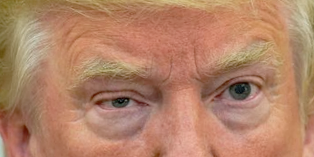
Superior tarsal muscle helps raise the upper eyelid and has a sympathetic nerve supply. Denervation of this muscle causes ptosis which is milder compared to oculomotor (CN III) palsy which supplies the levator palpebrae superioris. Superior tarsal muscle is responsible for keeping upper eyelid in a raised position after levator palpebrae superioris raises it. This explains the partial ptosis seen in Horner syndrome. The lower eyelid may be slightly elevated owing to denervation of lower lid muscle which is analogous to the superior tarsal muscle.
Sympathetic nervous supply is responsible for the dilation of the pupil (mydriasis). When disrupted, parasympathetic supply is uninhibited, and constriction of the pupil (miosis) ensues. The reaction of the pupils to light and accommodation is normal as those systems do not depend on sympathetic nerve supply.
Ipsilateral anhidrosis, another classic presentation, depends on the level of interruption of sympathetic supply. Anhidrosis with first-order neuron lesions affects the ipsilateral side of the body as the sympathetic supply from its central origin. The ipsilateral face is involved in lesions involving the second-order neurons. Postganglionic third-order neuron lesions occurring after the vasomotor and sudomotor fibers have branched off show very limited involvement of the face (area adjacent to ipsilateral brow).
Iris heterochromia (relevant deficiency of pigment in the iris on the affected side) is seen in children younger than 2 years and in the congenital form of Horner syndrome. [6]
History and Physical
Localization of the lesion in Horner syndrome is crucial in subsequent management. A detailed history and physical examination are, therefore, of vital importance. When evaluating, the following points need to be considered:
- Balance, hearing, sensory, and swallowing problems can point towards a more central process involving the first-order neurons
- Prior history of trauma or surgical intervention involving head, face, neck, shoulder, or back point towards involvement of second-order neurons
- A detailed past medical and medication history to rule out the use of a miotic or mydriatic agent
- A headache, double vision, facial numbness, or pain indicate third-order neuron involvement
- The presence of anhidrosis and its location can help in localization
- A detailed history of a headache if present
- Longstanding symptoms point towards a more benign underlying cause versus recently progressive symptoms such as weight loss, hemoptysis, low-grade fever, and lymphadenopathy
- Facial flushing suggests a preganglionic lesion
- Facial or orbital pain in combination with miosis and ptosis should point towards Raeder paratrigeminal syndrome
- History of skin lesions or previously diagnosed herpes zoster infection
- Severity and location of pain
A detailed examination of the eyes is warranted and should include the following:
- Reactivity of the pupils to light and accommodation
- Measurement of pupillary diameter in dim and bright light
- Examination of upper eyelid for ptosis and fatigability
- Evaluation of extraocular movements
- Vision, including color vision and visual fields
- Slit lamp examination for structural details
- Evaluation of the presence of nystagmus
A detailed exam can reveal a round and constricted pupil. The affected pupil exhibits dilation lag (dilates more slowly), and the unequal pupils are appreciated more in darkness compared to light. The ciliospinal reflex may be absent.
Furthermore, partial ptosis, iris heterochromia, apparent enophthalmos, contralateral eyelid retraction, injected conjunctivae, and no change or transient decrease in intraocular pressure may be seen. [14][15][16]
It is important to perform a detailed systemic examination, paying specific attention to neurological, pulmonary, and cardiovascular systems and considering various differentials discussed later.
Evaluation
Labs
- Initial workup should include complete blood count (CBC), erythrocyte sedimentation rate (ESR), and serum chemistry panel
- Urine or blood cultures may be ordered if an infectious agent is suspected.
- Testing for suspected neurosyphilis, HIV along with thyroid function, vitamin B-12, and folate levels may be ordered if indicated following detailed history and examination.
- Urine testing for elevated metabolic catecholamine by-products is important in the pediatric population with suspected neuroblastoma.
- Purified protein derivative (PPD) is warranted in suspected tuberculosis.
Imaging
- A chest X-ray followed by computed tomography (CT) scan must be ordered in patients when pulmonary malignancy is suspected.
- Head CT and magnetic resonance imaging (MRI) are advised in cases of possible stroke.
- MRI is warranted and preferred over ultrasonography when carotid artery dissection is a possibility (painful Horner syndrome). [17]
Pharmacological Testing
This is the most helpful testing modality for diagnostic localization. [18]
Topical cocaine test
Cocaine acts an indirect sympathomimetic inhibiting the reuptake of norepinephrine from the synaptic cleft. Cocaine solution (ranging from 2% to 10%) is instilled into both eyes. Both eyes are evaluated after at least 30 or more minutes for an optimal response. Denervation in the affected eye causes it to dilate poorly compared to the normal one. Anisocoria of 0.8 mm or more is considered diagnostic. The test does not help in identifying the level of lesion.
The test has other disadvantages such as comparatively high prices of the compound, time-consuming, test yielding ambiguous results, and the metabolites of cocaine can be detected in urine. [19]
Topical hydroxyamphetamine test
This test is particularly helpful in the localization of the lesion. Hydroxyamphetamine stimulates the release of stored norepinephrine from the postganglionic terminals into the synapse. Postganglionic third-order lesions can be differentiated from presynaptic second-order or first-order ones.
Two drops of 1% hydroxyamphetamine solution are instilled into both eyes. The affected eye (third-order lesion) will not dilate as well as the normal eye. While in the case of intact postganglionic fibers (first and second-order lesions), the affected pupil dilates to an equal or greater extent.
Disadvantages involving this test include it not being able to be performed on the same day as the cocaine test and false-negative results. [19]
Topical apraclonidine test
This test is considered the test of choice due to good sensitivity and overall practicality. Apraclonidine acts as a weak alpha1-agonist and strong alpha2-agonist. It is categorized as an ocular hypotensive agent. The upregulation of alpha1-receptors in Horner syndrome translates into an exaggerated response of the iris dilators (denervation supersensitivity) to an agonist agent like apraclonidine.
A 0.5-1% solution is instilled in both eyes. The affected eye will show mydriasis, while the normal eye is predominantly insensitive. Consequent instillation of the solution results in evident reversal of anisocoria (the affected pupil dilates and the normal pupil constricts). This is because of the stronger alpha 2 agonist activity of compared to the weaker alpha 1 agonist activity of apraclonidine.
Some disadvantages of this form of testing include false-negative results in acute cases, systemic side effects in the pediatric population, inability to localize lesion, and relatively long half-life of the drug.
Other agents have been proposed but fall short of clinical relevance due to several reasons.
- Pholedrine, an N-methyl derivative of hydroxyamphetamine, is roughly half as potent as hydroxyamphetamine and has shown comparable results but due to limited availability, has been restricted.
- Adrenaline, a direct-acting sympathomimetic, also has been proposed but has poor penetration through cornea and sensitivity to the drug is variable.
- Phenylephrine, a selective alpha1 agonist, also has been proposed but suffers from poor penetration through the cornea and varying results, depending on a degree in denervation. [20]
Treatment / Management
Treatment options are based on the diagnosis and management of the underlying cause. Timely diagnosis is of critical importance followed by referral to an appropriate specialist. Healthcare professionals are advised to incorporate the importance of eye examinations into their acumen.
Differential Diagnosis
As previously described, pain accompanied with Horner syndrome points towards a more insidious underlying cause and should be evaluated thoroughly. Systemic symptoms such as weight loss and progressive fatigue can indicate an underlying malignancy. Furthermore, Horner syndrome can be an early manifestation of neuroblastoma in the pediatric population.
Carotid artery dissection can present with a unilateral headache and facial or neck pain. If suspected, urgent appropriate workup and treatment are warranted.
Raeder paratrigeminal syndrome also can present with headaches but is accompanied by trigeminal nerve (CN V) impairment.
Anisocoria and/or ptosis can be due to myriad diseases and conditions such as Holmes-Adie syndrome, neurosyphilis (Argyll Robertson pupil), third nerve palsy, or optic neuritis.
Enhancing Healthcare Team Outcomes
There are many causes of Horner syndrome and thus the condition is best managed by an interprofessional team that includes nurse practitioners. The key is to determine the location of the eye defect. Thus, it is important to perform a detailed systemic examination, paying specific attention to neurological, pulmonary, and cardiovascular systems and considering various differentials discussed later.
The outcomes depend on the cause.
(Click Image to Enlarge)
Horner syndrome
Image courtesy S Bhimji MD