Ichthyosis X-Linked
- Article Author:
- Jonathan Crane
- Article Editor:
- Amy Paller
- Updated:
- 9/22/2020 10:45:01 PM
- For CME on this topic:
- Ichthyosis X-Linked CME
- PubMed Link:
- Ichthyosis X-Linked
Introduction
X-linked ichthyosis (XLI), known as steroid sulfatase (STS) deficiency and X-linked recessive ichthyosis, is a genetic skin disorder recognized in 1965 by Drs. Wells and Kerr. Because of the abnormal shedding, skin tends to be dry and accumulates polyg
- Acrodermatitis enteropathica
- Acute complications of sarcoidosis
- Acute retinal necrosis
- Atopic dermatitis in emergency medicine
- Herpes zoster
- Interstitial keratitis
- Opthalmologic manifestations of leukemia
- Ocular manifestation of HIV
- Psoriasis
- Retinitis pigmentosa
onal scales.[1]
Etiology
A deficiency in STS (Xp22.3) is responsible for the abnormal cutaneous scaling seen in X-linked ichthyosis. Most X-linked ichthyosis patients have extensive deletions (complete or partial) of the STS gene; however, point mutations may result in complete STS deficiency. Female carriers of STS gene do not exhibit any manifestations because the gene is localized to a region of the X-chromosome that does not undergo X-inactivation. De novo STS mutation also occurs.[2][3]
Epidemiology
X-linked ichthyosis is the second most common form of ichthyosis, with ichthyosis vulgaris the most common type. X-linked ichthyosis is equally reported in all ethnic groups and races worldwide. As the name of the disease suggests, X-linked ichthyosis almost exclusively affects males. The incidence of X-linked ichthyosis is reported as 1 out of 2500 to 1 out of 6000 males. Three X-linked ichthyosis female cases have been described, all of these patients were offspring of an affected father and a carrier mother.[2][4]
Pathophysiology
Absent STS activity in the epidermis causes retention hyperkeratosis. STS hydrolyzes steroid sulfates to their unconjugated (unsulfated) forms and participates in the regulation of barrier permeability and desquamation. The absence of STS activity results in the accumulation of cholesterol sulfate in the stratum corneum, leading to corneocyte cohesion, hyperkeratosis, and impaired skin permeability. A deficiency or absence of STS may be associated with asymptomatic corneal opacities and rarely cryptorchidism. Abnormalities in neurologic and cognitive development and/or anosmia have been described when the deletion encompasses more than one gene (contiguous gene syndrome). [4]
Histopathology
The histologic features of X-linked ichthyosis include subtle hyperkeratosis with dermal perivascular inflammation. Notable changes can be appreciated in the stratum corneum and granular layers of the epidermis. Moreover, the granular layer can appear normal or thin or may be absent.[5]
History and Physical
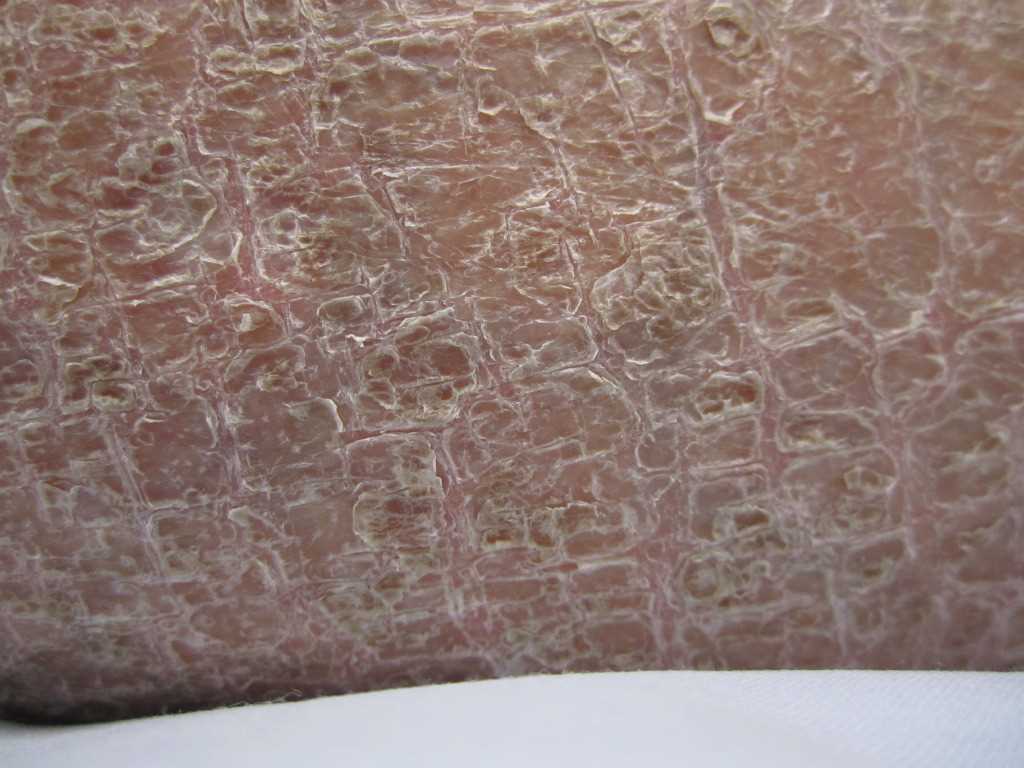
Skin findings usually appear within the first year of life, and 15% to 20% have manifestations at birth. In general, scaling and erythema are seen if at birth, but collodion baby has been described. Most others show the scaling with mild to no erythema in infancy. Typical clinical findings include mild, diffuse scaling that develops over time, mild desquamation with larger polygonal scales affecting mainly the scalp, anterior aspects of the lower extremities (shin), and other extensor surfaces. The flexures (popliteal and antecubital fossae), palms, and soles are spared, as are the hair and nails. The scales tend to increase throughout childhood and continue into adolescence. Pruritus usually is absent. Desquamation is typically mild during the summer months and is exacerbated by dry and cold weather. The differential diagnosis is ichthyosis vulgaris, which does not tend to have its onset during the first 3 months (although associated atopic dermatitis may occur early). Mild lamellar ichthyosis can appear similar. Asymptomatic corneal opacities are the most common eye finding (present in up to 50% of affected males and 25% of female carriers). Cryptorchidism has been reported to affect up to 20% of X-linked ichthyosis patients, but in practice, it is uncommonly found. There have not been adverse effects on sexual development, testosterone levels, or fertility.[6][7][8]
Evaluation
The diagnosis of X-linked ichthyosis is based on biochemical and/or genetic analysis. The most accurate diagnostic method is genetic analysis. Chromosomal microarray will often detect the STS deletion as well as contiguous gene syndrome. STS hydrolyzes sulfated (conjugated) alkyl steroid sulfates, including dehydroepiandrosterone (DHEAS), to their unsulfated (unconjugated) form (DHEAS->DHEA). As a result, decreased STS activity results in higher conjugated and lower unconjugated steroid sulfates. Notably, in women carrying an affected fetus, absent placental STS activity will lower the mother’s unconjugated serum estriol (uE3). A falsely low uE3 level will result in false-positives in second-trimester Down syndrome screening tests; therefore, X-linked ichthyosis may be diagnosed prenatally as an unexpected finding in women undergoing elective genetic screening tests during the second trimester of pregnancy. Fluorescence in situ hybridization (FISH) analysis or chromosomal microarray analysis can be used to confirm the diagnosis; affected boys will have variable (and sometimes no) manifestations, presenting during infancy. For those who carry microdeletion within the STS gene, assaying for STS activity from skin fibroblasts, keratinocytes, or lymphocytes of X-linked ichthyosis-suspect patients is recommended. For carrier detection, both multiplex quantitative fluorescent PCR (QF-PCR) and fluorescent in situ hybridization (FISH) are capable of detecting complete deletion of the STS gene and identifying a female carrier.[9][10][11][12]
Treatment / Management
The medical management of X-linked ichthyosis is directed at reducing scales, decreasing skin dryness, and improving skin appearance. This can be accomplished with regular bathing, and use of emollients and keratolytic agents. Petrolatum or humectant-based moisturizers should be applied to damp skin. Topical keratolytics are probably best avoided in the first 6 months of life and should be used with caution if over large body surfaces In children, given the risk of systemic absorption and its associated toxicities. In severe forms of X-linked ichthyosis, patients may benefit from the intermittent use of topical or even systemic retinoids. Substantial evidence on the efficacy of keratolytic, topical, and systemic retinoids for the treatment of mild to severe X-linked ichthyosis is limited. Patients clinically respond well to 10% to 20% urea cream, lactic acid and/or salicylic acid-containing moisturizers. Tazarotene, 0.05% gel, has been shown to cause marked clinical improvement in X-linked ichthyosis, while oral acitretin induced dramatic improvement in scaling and erythema. Ophthalmology examination is generally unnecessary for affected males since the punctuate corneal opacities are asymptomatic. If cryptorchidism is present, a urology consult is warranted, followed by routine monitoring for testicular carcinoma, which has only been reported in a single patient and was unrelated to cryptorchidism.[13][14][15]
Differential Diagnosis
- Acrodermatitis enteropathica
- Acute complications of sarcoidosis
- Acute retinal necrosis
- An ocular manifestation of HIV
- Atopic dermatitis in emergency medicine
- Herpes zoster
- Interstitial keratitis
- Ophthalmologic manifestations of leukaemia
- Psoriasis
- Retinitis pigmentosa
Pearls and Other Issues
The most common cognitive and behavioral disorders in X-linked ichthyosis patients include attention-deficit hyperactivity disorder (ADHD, inattentive subtype), and autistic-spectrum disorder (language/communication difficulty), but there is not good evidence that these are more common in X-linked ichthyosis than in control male population. Contiguous gene syndrome from contiguous gene deletions may variably present with retardation, hypogonadotropic hypogonadism and/or anosmia; the latter two features are seen in Kallmann syndrome. [13][14][15][16]
Enhancing Healthcare Team Outcomes
Best results in the management of X-linked ichthyosis requires a coordinated interprofessional team of specialty trained clinicians and nurses to assist in patient education and coordination of care. [Level V]

