Fungal Keratitis
- Article Author:
- Gabriel Castano
- Article Author:
- Ayman Elnahry
- Article Editor:
- Pradeep Kumar Mada
- Updated:
- 8/10/2020 9:28:58 PM
- For CME on this topic:
- Fungal Keratitis CME
- PubMed Link:
- Fungal Keratitis
Introduction
Bacteria, viruses, fungi, and protozoa are all causes of microbial keratitis. Fungal keratitis (FK) was first documented in 1879, and its incidence has been increasing for the past 30 years. It accounts for about 40% to 50% of all microbial keratitis cases.[1][2][1] FK is a serious condition that if not properly treated can lead to corneal destruction and endophthalmitis with severe loss of vision. Early diagnosis and management are, therefore, essential to prevent long-term complications including blindness.[3][4]
More than 100 different fungal species are capable of causing FK.[5] Monomorphic fungi can be classified into yeast and filamentous fungi and both are responsible for cases of FK. The specific type of fungus responsible for FK depends on several factors including personal risk factors, regional temperature, climate conditions, geographic location, and urbanization.[6]
Trauma, immunocompromised state, ocular surface disease, and contact lens wear are the most prevalent personal risk factors associated with FK and may predispose to different types of fungal infections.[6][7][6]
Clinically, it may be challenging to diagnose a fungal infection as a cause for keratitis and diagnostic delays are common with negative or delayed culture results. A high index of suspicion is, therefore, usually required depending on the presence of specific risk factors for FK. Even if the diagnosis is made, management is still challenging as many antifungal agents have poor penetration into the cornea.
Etiology
Many fungal species can result in FK, however, the most commonly implicated are Fusarium, Aspergillus, and Candida species.[5] The causative organism of FK may differ according to several factors including regional temperature, climate, and urbanization. In a study performed in India on FK, Aspergillus species were the most commonly isolated species followed by Fusarium.[8] This was also confirmed in several other studies from India, with one study showing that Aspergillus was responsible for more than 55% of all FK cases, suggesting that Aspergillus species are the most common cause of FK in the Indian subcontinent.[9] Other studies, however, have shown that Fusarium was the most common etiology of FK in other parts of the world including south India.[10][11] In a large study from the USA over a 7 year period, 39% of FK cases were caused by Fusarium, while 22% were caused by yeasts including Candida.[7] In another study of 2065 cases of confirmed fungal keratitis in central China, the predominant fungal species was also Fusarium (>50%), followed by Aspergillus (>9%) and Alternaria (>7%).[12] This was similar to findings from a study performed in north China.[1] In a study from the United Kingdom, however, Candida (57%) was the most prevalent causative fungus followed by Aspergillus (17%).[6] This indicates that the causative organism of FK may differ according to the geographic location.
The prevalence of specific types of fungi causing FK also depends on specific patient-related risk factors. For example, Fusarium species are found more commonly in association with trauma and contact lens use,[7][13][7] while Candida species are found more in association with ocular surface disease and topical steroid use.[6] Trauma, especially with vegetative matter, is an important risk factor for filamentary fungal infections in general, including Fusarium, however, it may also result in bacterial infections.[14][12][14] Accordingly, certain occupations, like those involving agriculture, may carry an increased risk of microbial keratitis including fungal keratitis.[12] The risk of corneal fungal infections may also be increased with a lack of personal hygiene including poor hand hygiene and overnight contact lens wear.[15]
Corneal refractive surgery including laser in situ keratomileusis (LASIK) may also rarely be a cause of fungal keratitis that could lead to severe consequences if not properly treated.[16] Various fungal species responsible for post-LASIK FK have been isolated including Candida, Fusarium, Aspergillus, and Alternaria species.[17][18][19][20] This could be due to inappropriate operative room sterilization techniques or poor postoperative hygiene.[17]
Epidemiology
Like most infectious diseases, geographical location, and socioeconomic status influence the prevalence and cause of FK. In the United States, warm places in southern locations like Florida have a higher incidence of FK compared to colder places in the north.[21][22][21] Fusarium, Candida, and Aspergillus are the most frequently isolated species causing FK in the USA,[7] while in India, Aspergillus is the most common cause.[9] Fusarium is a particularly common cause of FK in warm climates such as Brazil,[23] while Candida may be more common in temperate climates.[22]
The overall incidence of FK seems to be markedly higher in certain parts of the world. In a study of FK in Hyderabad India, 1360 patients with FK were seen over a period of 10 years in a single institute, while in a study from central China, 2065 cases were documented over a 9 year period.[12] In a study in Melbourne Australia, however, only 56 cases were seen in 8 years,[24] while in a study from New York, only 61 eyes with FK were documented over a 16 year period.[25] This may be due to climate, environmental, occupational, or behavioral differences.
An estimated 45 million residents in the USA use contact lenses.[26] Contact lens use can lead to microbial keratitis, especially if not properly used, and it is estimated that more than 80% of contact lens users in the USA in different age groups have at least one behavior that makes them at risk for developing a contact lens-associated eye infection.[27] These risky behaviors include wearing contact lenses overnight and washing contact lenses with tap water. In a major outbreak of Fusarium keratitis across the USA and many other countries of the world between 2005 and 2006, however, cases were eventually traced back to a specific brand of multipurpose contact lens disinfecting solution, indicating the importance of the continuous monitoring of these products and their components to decrease the risk of future outbreaks of contact lens-associated eye infections.[28]
Several studies have shown a higher incidence of FK in young adult males, possibly due to more outdoor activities and higher incidence of trauma.[29] In a large study of Chinese patients with FK, more than 60% were males, and more than 75% were aged between 30 and 60 years.[12]
Pathophysiology
FK commonly results from fungal access into the corneal stroma through a defect in the corneal epithelium; this is probably the reason behind the increased risk of FK in association with trauma and foreign bodies that lead to loss of the integrity of the protecting epithelium. Injury with vegetative matter may result in direct inoculation of fungal conidia present on their surface into the corneal stroma leading to initiation of infection or injure the overlying corneal epithelium permitting fungal invasion.[1][13] Once in the tissue, fungi start to replicate in the stroma spreading circumferentially with satellite lesions and penetrating deeply into the stroma to reach Descemet's membrane and eventually the anterior chamber. This may eventually lead to corneal perforation and endophthalmitis if the infection is not overcome by the natural body defenses or if proper treatment is not administered. Fungi can also spread from the cornea into the sclera and surrounding structures, leading to severe consequences such as scleritis and panophthalmitis.
The cornea is avascular, and on top of its barrier capacity, is immune privileged with restricted defense mechanisms, dendritic cells, immune cells, and immunoglobulins, making it easy to be colonized by fungi. Fungi also secrete various toxins and enzymes including serine proteases and matrix metalloproteinases that help in corneal invasion and colonization.[2] The associated inflammatory reaction from immune cells in the cornea, especially polymorphonuclear leukocytes, may result in further damage to corneal tissue.[2]
Histopathology
Gross granular infiltration of the corneal epithelium and the anterior stroma is the main finding in FK with collagen destruction, coagulative necrosis, and stromal fungal infiltration seen on microscopy. FK is usually associated with less purulent inflammatory cellular infiltration compared to bacterial keratitis.[30] Having low neutrophil infiltration in the cornea is a positive finding as they are the ones that contribute the most to the destruction of the cornea with the intent to control the causative organism.[2] Lymphocytes and plasma cells are also often seen in FK.
Various stains can be used to detect fungi in corneal tissue or scrapings including potassium-hydroxide, gram staining, Giemsa staining, lactophenol cotton blue, methenamine silver, and calcofluor white.[22] This allows visualization of both fungal hyphae and yeast cells and helps in the early differential diagnosis of the causative organism before the results of cultures are available.[31] It may also allow the detection of a mixed fungal and bacterial infection. Different hyphal growth patterns have been observed histopathologically and may also help in the differential diagnosis of the specific filamentous fungus responsible for the condition including Fusarium hyphae that lay parallel to the lamellae of the corneal stroma, and Aspergillus hyphae that grow vertically.[32]
History and Physical
The history and clinical picture of FK may differ according to the causative organism, depending on whether it is a filamentous fungus or yeast.[22] General features of microbial keratitis are usually present including blurring of vision, pain, redness, discharge, and blepharospasm. In addition to corneal infiltration, patients may have signs of inflammation of the anterior chamber with hypopyon. In cases of FK due to filamentous fungi, there may be a history of trauma with vegetative matter, and patients usually present with an ulcer characterized by elevated firm slough, hyphate lines that extend beyond the edge of the central ulcer, and feathery satellite stromal lesions. Patients with FK due to yeast fungi may have a history of an ocular surface disease or immunosuppression with a clinical picture that resembles more bacterial keratitis but with a slower progression. Although it may sometimes be possible to differentiate a fungal etiology from a bacterial etiology of microbial keratitis based on the clinical picture alone, this may not always be the case, and prediction of the causative fungal genus or species responsible for the condition may be even more challenging and inaccurate.[33]
Evaluation
Laboratory evaluation of a case of FK begins with the collection of an appropriate sample for testing. These specimens are used for direct examination by microscopy using various stains such as Giemsa and Gomori methenamine silver stains, culture, histologic testing, and other tests.[34] Because fungi have the predilection to penetrate deep into the cornea, tissue swabbing is usually inadequate in confirming a fungal infection and deep corneal scrapings are usually needed.[35][36] A corneal biopsy may sometimes be needed to obtain an adequate sample. Ideally, every sample should be sent for polymerase chain reaction (PCR) and culture, especially if corneal scraping stains are negative.[35][36] Fungal cultures usually take from 1 to 35 days for fungal growth and include Sabouraud glucose neopeptone agar, blood agar, thioglycolate broth, and brain heart infusion, which allow isolation of different types of fungi.[34][36] A major disadvantage of fungal cultures, however, is the relatively low sensitivity with false-negative results possibly due to the small amount of material available from corneal scrapings.[35][36] This is especially true for cases that have been treated with antifungal agents. PCR testing has been proposed as a more sensitive method of diagnosing FK that allows accurate species identification and takes about 4 to 8 hours, however, it may be less specific with false-positive results possibly due to the amplification of non-pathogenic organisms in the sample.[22][34][36] Targets used for PCR amplification include fungal 18S rRNA and 28S rRNA. PCR can be performed from a small sample of ocular tissues or fluids like tears or aqueous humor but requires very specialized equipment that is expensive and may not always be available.
Beta-D-glucan is a component of the cell wall of many fungal species that can be detected in blood samples of patients with invasive fungal infections.[37] Previously, it has been shown that beta-d-glucan testing is a more rapid and more sensitive method of diagnosing systemic fungal infections than fungal cultures especially in organisms that do not grow in blood cultures such as Aspergillosis.[38] Beta-D-glucan may be detected in tear samples of patients with fungal keratitis facilitating early and rapid diagnosis.[39] It can also be detected in intraocular fluids of cases complicated by fungal endophthalmitis.[40]
Non-invasive methods, including confocal microscopy and anterior segment optical coherence tomography (AS-OCT), can also be used to detect microorganisms responsible for microbial keratitis, including fungi, using in vivo examination techniques. Confocal microscopy may allow detection of hyphae-like branching white lines in the area of infiltration in cases of filamentous fungi,[41] while AS-OCT may show early localized and diffuse areas of necrotic stromal cystic spaces in cases of infection by Aspergillus species.[42] Both modalities can also be used in the follow up of the response to treatment.[43]
Ophthalmic B-scan ultrasonography can be useful in the diagnosis and follow up of cases suspected of developing endophthalmitis especially if corneal fungal infiltration precludes examination with ophthalmoscopy.
Treatment / Management
Medical Treatment
All the currently available antifungal agents are fungistatic but not fungicidal, which necessitates a prolonged period of treatment until the body defenses can completely eradicate the fungal organism. Topical natamycin 5% is usually the first treatment of choice for FK especially filamentous fungi,[44][45] although primary treatment failure has previously been reported in 31.3% of cases.[46]
Other topical agents used in the treatment of FK include amphotericin B 0.15-0.3%, voriconazole 1%, econazole 1%, itraconazole 1%, and miconazole 1%. Amphotericin B is the preferred treatment of choice for yeasts. Voriconazole 1% has better penetration into the eye and is purported to be a superior alternative to natamycin. It can also be used as intrastromal or intracameral injections.[47]
Subconjunctival injections of antifungal agents such as miconazole and fluconazole may be used in patients with poor compliance and those with severe keratitis.
Systemic treatment can also be used in the form of intravenous amphotericin B or oral itraconazole or fluconazole.
Decisions to continue therapy as based on the following biomicroscopic findings:
- A decrease in pain and size of infiltrate
- A decrease in satellite lesions
- A decrease in the density of suppuration
- Blunting of the infiltrate edges
- A decrease in inflammation in the anterior chamber
- Continued reepithelialization
Recently published clinical trials have reported equal or inferior efficacy of topical 1% voriconazole (reconstituted from injection vial) compared with topical 5% natamycin eye drops in FK especially in that caused by Fusarium. These results, however, are contradictory to experimental and in vitro data. Sharma et al conducted a study of 118 patients where 58 patients were treated with voriconazole and 60 patients with natamycin. Despite the frequency of healed or resolving ulcers being similar on day 7 (natamycin 35/54, 65%; voriconazole 34/50, 68%), at the final visit the percentage of patients who had healed corneal ulcer were significantly higher in the group treated with natamycin (50/56, 89.2% versus 34/51, 66.6%; p=0.005).[48]
A Cochrane Database systematic review of the medical treatment of fungal keratitis was performed in 2015 and also concluded that natamycin may be more effective than voriconazole in the treatment of fungal keratitis but that also most studies were underpowered with variable quality.[49] The randomized controlled trials analyzed included many comparisons including topical 5% natamycin versus topical 1% voriconazole, topical 1% voriconazole vs intrastromal voriconazole, and topical 1% natamycin versus topical 2% econazole.
Surgical Treatment
Patients who fail to respond to medical therapy may require surgical intervention, including therapeutic penetrating and lamellar keratoplasty.[12][50][12] Therapeutic keratoplasty, however, can be associated with complications including recurrence of infection, endophthalmitis, and graft rejection.[50]
A recently available treatment modality is corneal collagen cross-linking which may sometimes be useful in medically resistant corneal ulcers or even in some early cases of fungal infection.[51][52][53][52] A recent randomized controlled clinical trial, however, did not show any benefit of this modality in fungal keratitis with an increased incidence of complications.[54] Conjunctival flaps can also rarely be used but may increase the incidence of graft rejection following keratoplasty due to corneal vascularization.[12]
If a corneal perforation occurs, a tectonic or therapeutic keratoplasty is usually required to save the eye. Cases complicated by endophthalmitis may require intravitreal injections of antifungal agents or even pars plana vitrectomy which can be performed using endoscopy in cases with poor posterior segment visualization due to extensive corneal infiltration.[55][56][57] Enucleation, however, may eventually be the last resort in a blind and painful eye with uncontrollable inflammation.
Healing of FK may result in central corneal scarring and opacification which may require penetrating or lamellar keratoplasty for the restoration of visual acuity.
Differential Diagnosis
- Bacterial keratitis
- Acanthamoeba keratitis
- Necrotizing herpetic keratitis
Prognosis
The prognosis depends on the causative organism, depth and extent of the infection, development of complications, and timing of treatment initiation. Some patients can be cured microbiologically by topical antifungals alone, but in some patients, therapeutic keratoplasty may be needed. The Descemet membrane, an interior basement membrane near the aqueous humor, is usually impermeable to bacteria but can be breached by fungal hyphae, leading to endophthalmitis; endophthalmitis is a rare consequence of fungal keratitis that has a poor prognosis. About 30% of fungal infections fail to respond to drug therapy and/or result in corneal perforation. Successful healing of FK usually results in central corneal scarring and opacification which may require penetrating or lamellar keratoplasty for the restoration of visual acuity.
Complications
- Corneal perforation
- Corneal melting
- Corneal scarring
- Scleritis
- Endophthalmitis
- Panophthalmitis
- Permanent blindness
Deterrence and Patient Education
Patients should be educated about the proper use of contact lenses and their cleaning products in order to decrease the risk of contact-lens associated corneal infections. Proper hygienic measures should be emphasized including frequent hand washing especially before handling contact lenses and avoiding overnight contact lens wear.
Pearls and Other Issues
- Fusarium, Candida, and Aspergillus species are the most common isolates in fungal keratitis worldwide.
- Elevated edges, branching ulcers, feathery margins, rough texture, and satellite lesions are features suggestive of fungal keratitis.
- The diagnosis is by corneal scrapings. Samples should be sent for PCR and cultures.
- Natamycin eye drops are frequently initially used.
Enhancing Healthcare Team Outcomes
The majority of patients with fungal keratitis present to the emergency department or their primary care provider; in order to expedite the referral process to an ophthalmologist, an interprofessional approach in diagnosis and care is vital. Any delay can lead to vision loss and permanent blindness. The majority of patients with fungal keratitis are managed as outpatients and treated with antifungal therapy for at least 12-16 weeks.
Patient education is vital in the prevention of fungal keratitis. Contact lens wearers need to be educated about hand washing, use of appropriate cleaning solution, and avoiding sleeping and swimming while wearing contact lenses. Contact lens wearers should also regularly follow up with the optician or ophthalmologist and get their eyes checked. All contact lens wearers should be informed of symptoms of eye infections and the need to immediately see an ophthalmologist if they experience pain, redness, or loss of vision. Ophthalmology nurses should educate patients, arrange for follow up, and ensure communication between the team members. Pharmacists should review medication dosages, check for drug interactions, and review methods of administration with patients.
Close follow up during treatment is required to ensure that the condition is not worsening. The eventual outcome depends on factors like overall patient health, the status of the immune system, and other comorbidities. Patients with a mild infection who are promptly treated have a good outcome, but in patients with an infection that has spread into the sclera, the prognosis is guarded. Data indicate that at least 30% of patients with fungal keratitis develop corneal perforation or fail to respond to drug therapy.[58][59] The evidence regarding the management of FK varies and ranges from large randomized controlled clinical trials with clear-cut results in certain aspects of its management (Level 1 evidence) to case series and expert opinion in other aspects (Level 5 evidence).
(Click Image to Enlarge)
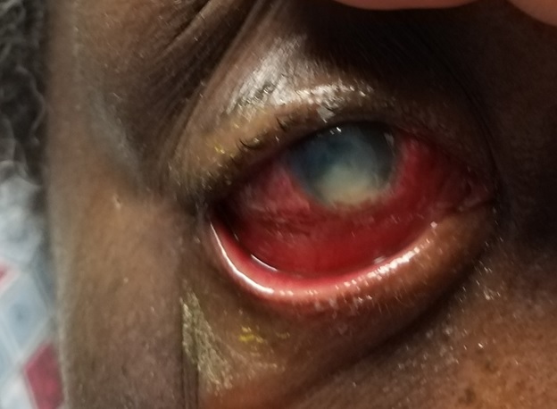
Fungal keratitis with injection,kemosis and hypopyon
Contributed by Gabriel Castano, MD