Lipoma
- Article Author:
- Logan Kolb
- Article Author:
- Siva Naga Yarrarapu
- Article Author:
- Muhammad Atif Ameer
- Article Editor:
- Juan Rosario-Collazo
- Updated:
- 10/6/2020 2:49:01 PM
- For CME on this topic:
- Lipoma CME
- PubMed Link:
- Lipoma
Introduction
Lipomas are benign subcutaneous tumors of fat cells (adipocytes) that present as soft, painless nodules that are most commonly seen on the trunk. [1][2][3] Lipomas usually range from 1- >10 cm. They are mesenchymal tumors and are found anywhere in the body where normal fat cells are present and are enclosed in a fibrous capsule. They are benign and have many histologic subtypes. The presence of multiple lipomas may be the presenting feature of a variety of syndromes.[4] Although lipomas are present in subcutaneous planes rarely, they may also involve fascia or deeper muscular planes.
Etiology
Lipomas represent the most common mesenchymal tumors of the human body. About 1 in every thousand-persons will have lipoma at some point in time. The majority of the lipomas affect the upper extremity, but they can be anywhere on the body where adipocytes are generally present. The precise cause of lipomas is unknown. A potential link between the trauma and lipoma formation is explained by many. It is also being speculated that trauma induces cytokines to release triggers preadipocyte differentiation and maturation.[5]
Genetics appear to play a role since 2% to 3% of affected patients have multiple lesions inherited in a familial pattern.[6] An association of gene association with chromosome 12 has been established in the solitary lipomas due to mutation in the HMGA2-LPP fusion gene.[7] There also are several genetic syndromes that feature lipomas as a clinical manifestation. The incidence of lipomas is increased in patients with obesity, hyperlipidemia, and diabetes mellitus.
Epidemiology
Lipomas have a slightly higher incidence in males compared to females. Although they can occur at any age, they are often noted between the fourth to sixth decades of life. Despite being one of the most common neoplasms in humans, the incidence and prevalence of lipomas have not been reported in the literature.
Pathophysiology
The exact pathophysiology of lipomas is unclear. However, benign lipomas can be present in almost any organ of the body. Subcutaneous lipomas are not usually fixed to the underlying fascia. That's why the fibrous capsule should be removed completely to prevent ist reoccurrence.
In the gastrointestinal (GI) tract, the lipomas present as submucosal fatty tumors. The most common locations include small intestines, stomach, and esophagus. Gastrointestinal lipomas become symptomatic only after luminal abstraction or bleeding. Mucosal erosion over lipoma may cause severe bleeding in the bowel.[8] Small bowel lipomas mostly occur in the elderly population and most commonly present in the ileum. They are usually pedunculated, submucosal lesions, and can obstruct the lumen of the bowel. [9][10][9] They can cause obstructive jaundice, pain, gastric outlet obstruction, and intussusception in younger patients.
Colonic lipomas are usually discovered incidentally on colonoscopy. A biopsy specimen of the lipoma will most likely reveal mature adipocyte, which is called the "naked fat' sign. As with other lipomas, colonic lipomas can also cause pain with obstruction and intussusception.
However, several cytogenetic abnormalities have been identified, including the following:
- Mutations in chromosome 12q13-15, 65% of cases
- Deletions of 13q (10% of cases), rearrangements of 6p21-33, 5% of cases
- Unidentified mutations or normal karyotype, 15% to 20% of cases
Rearrangements of the 12q13-15 result in fusion of the high-mobility group AT-hook 2 (HMGA2) gene to a variety of transcription regulatory domains that promote tumorigenesis.[11][12]
Histopathology
Histologic examination of lipomas reveals mature, normal-appearing adipocytes with a small eccentric nucleus. Adipocytes are intermixed among thin fibrous septa containing blood vessels. These features are indistinguishable from adipocytes in the subcutaneous tissue. Histologic subtypes of lipomas include angiolipomas, myelolipomas, angiomyolipomas, myelolipomas, fibrolipomas, ossifying lipoma, hibernomas, spindle cell lipomas, pleomorphic lipomas, chondroid lipomas, and neural fibrolipomas. Common lipomas and its variants must be distinguished from liposarcomas, which are a malignant lipomatous neoplasm containing lipoblasts, which are characterized by coarse vacuoles and one or more scalloped, hyperchromatic nuclei.
History and Physical
Lipomas typically present as soft, solitary, painless, subcutaneous nodules that are mobile and not associated with epidermal change. A characteristic "slippage sign" may be elicited by gently sliding the fingers off the edge of the tumor. They are typically slow-growing and grow to a final stable size of 2 to 3 centimeters. However, they are occasionally greater than 10 centimeters and referred to as "giant lipomas." Lipomas may appear anywhere on the body but tend to favor the fatty areas of the trunk, neck, forearms, and proximal extremities. They are rarely seen in acral areas. Lipomas may affect many cutaneous and noncutaneous sites, including dermal, subcutaneous, and subfascial tissues along with intermuscular, intramuscular, synovial, bone, nervous, or retroperitoneal sites.
Multiple lipomas are present in 5% to 10% of affected patients and are usually associated with familial lipomatosis or numerous other genetic disorders described in the differential diagnosis section. The use of protease inhibitors in HIV patients may induce lipomas and lipodystrophy; therefore, a thorough past medical and medication history should be obtained.
Sign and symptoms of lipomas depend largely on the location and size of the lipomas. It can cause respiratory distress related to bronchial obstruction; patients may present with either endobronchial or parenchymal. Cardiac lipomas are located mainly subendocardial, are rarely found intramurally, and are normally unencapsulated; they appear as a yellow mass projecting into the cardiac chamber lesions. Patients with esophageal lipomas can present with obstruction, dysphagia, regurgitation, vomiting, and reflux; esophageal lipomas can be associated with aspiration and consecutive respiratory infections.
Evaluation
Common lipomas frequently are diagnosed clinically and sent for histologic examination after complete surgical excision.[13] Radiologic imaging before surgery may be prudent in cases featuring the following:
- Giant size (greater than 10 centimeters),
- Rapid growth
- Pain
- Fixation to underlying tissues
- Location in deep tissues, the thigh, or retroperitoneal space
For most subcutaneous lipomas, no imaging studies are required. However, lesions in the gastrointestinal (GI) tract may be visible on GI contrast studies, While imaging like ultrasound, MRI, and CT scans is required for lipomas an atypical location.[14] As lipomas are radiolucent, soft tissue radiography can be diagnostic, but it is only employed when the diagnosis is in doubt clinically.
Treatment / Management
The majority of the patients who seek treatment for lipomas is due to cosmetic reasons. Lipomas do not involute spontaneously, although dramatic weight loss may make lesions more clinically prominent. Stable lesions often are observed clinically. The decision to treat depends on numerous factors, including lesion size, anatomic location, symptoms such as pain, and patient comorbidities. If treatment is desired, surgical excision is commonly employed. Large lipomas have been removed via liposuction.[15][16][17]
Non-operative therapies like endoscopy and colonoscopy are utilized for the lipomas of the gastrointestinal tract (GI). Colonoscopic removal of lipomas is associated with a higher risk of colon perforation.
Complete surgical excision of the capsule is recommended. Intracardiac lipomas are usually lobulated, and all lobules must be removed for a better outcome. Subcutaneous lipomas are removed for cosmetic reasons. Therefore a cosmetically pleasing incision should be used. The incision is usually placed directly over the mass in a line of skin tension. Local removal is indicated in intestinal lipomas, causing obstruction and hemorrhage; the uncertainty of diagnosis for an intramural intestinal mass also warrants resection. If esophageal lipomas can not be endoscopically removed, surgical excision is indicated, whether by a transhiatal or a transthoracic approach.
Differential Diagnosis
The differential diagnosis includes but is not limited to epidermoid cysts, hibernomas, angiolipomas, angiomyolipomas, and liposarcomas. Epidermoid cysts typically feature a punctum at its surface; however, lesions lacking this feature may be indistinguishable from lipomas. Hibernomas are benign, rare, slow-growing masses of brown fat that typically occur in the mediastinum or interscapular region of the back and present as a 3 to 12 centimeter, slow-growing mass in an adult. Angiolipomas are often painful, less than 2 centimeters in width, well-circumscribed, and typically appearing on the forearm of teens or young adults. Angiomyolipomas often present in acral locations of adult males. Liposarcomas most commonly present as deep-seated tumors in the retroperitoneum or, classically, on the thighs.
Multiple lipomas can be a manifestation of the following syndromes:
- Proteus syndrome due to activating mutations in AKT1 oncogene (or occasionally PTEN mutations). It consists of lipomas, epidermal nevi, hemangiomas, palmoplantar cerebriform connective tissue nevi, hyperostoses of the epiphyses and skull, and scoliosis.
- Dercum disease (adiposis dolorosa) consists of multiple painful lipomas on the trunk and extremities, which often have overlying paresthesias of the skin. It usually affects postmenopausal women who suffer from psychiatric disorders.
- Familial multiple lipomatosis- patients typically present in the '30s with hundreds of encapsulated and noninfiltrating lipomas. It can be inherited in an autosomal dominant pattern.
- Benign symmetric lipomatosis (Madelung disease) involves diffuse, infiltrative, symmetric painless lipomatous growths affecting the head, neck, and shoulder region. Middle-aged alcoholic men are usually present with this syndrome. Mutations in the mitochondrial tRNA lysine gene have been identified in a few of the affected patients.
- Gardner syndrome is due to autosomal dominant mutations in the adenomatous polyposis coli (APC) gene. Almost all patients develop adenocarcinomas of the gastrointestinal tract. Cutaneous changes include multiple lipomas or fibromas. Other associated findings include congenital hypertrophy of pigment epithelium of retina, osteomas of the skull, maxilla and mandible, supernumerary teeth, and various malignancies, including papillary thyroid carcinomas, adrenal adenomas, and hepatoblastomas.
- Multiple endocrine neoplasia (MEN) type 1 is due to autosomal dominant mutations in the MEN1 gene. It consists of the parathyroid, pituitary, and pancreatic tumors. Dermatologic changes include multiple lipomas (which may also occur in visceral sites), collagenomas, angiofibromas, and cafe au lait macules.
- Cowden syndrome is due to mutations in the PTEN gene. It is associated with multiple lipomas, oral papillomas, facial trichilemmomas, punctate palmoplantar keratoses, and malignancies, including breast adenocarcinoma, thyroid follicular carcinoma (TFC), endometrial carcinomas, and hamartomatous polyps of the gastrointestinal tract.
- Bannayan-Riley-Ruvalcaba syndrome (BRRS) is also due to PTEN gene mutations and may represent a pediatric form of Cowden syndrome. Clinical findings include multiple lipomas, intestinal hamartomas, genital lentigines, macrocephaly, and mental retardation.
Prognosis
The prognosis is excellent for benign lipomas. Recurrence is not common but may develop if the excision was incomplete.
Complications
Gastrointestinal lipomas can cause obstructive symptoms and bleed secondary to ulceration. Cardiac lipomas may cause embolism if they interfere with the anatomy of the cardiac chamber. Although rare but local lipomas may cause nerve compression. As these lipomas enlarge, they can compress the adjacent nerves and local structure. The majority of lipomas have a benign course and don not recur after surgical removal. It is advisable to complete excise the capsule to prevent ist reoccurrence.
Consultations
Lipomatous lesions arising in the midline sacrococcygeal region can be a manifestation of spinal dysraphism (along with skin tags, hair tufts, angiomas, and a variety of other cutaneous changes). For this reason, a neurosurgery consultation is indicated for lipomatous lesions in this region.
Enhancing Healthcare Team Outcomes
Lipomas are commonly encountered by the primary care provider and nurse practitioner. However, the definitive management is usually done by a dermatologist, general surgeon, or a plastic surgeon. There are many ways to treat lipomas, but asymptomatic lesions should be left alone. Lipomas usually incur minimal morbidity- which in most cases is poor cosmesis.[18] an interprofessional team approach is best with a primary care provider, nurse practitioner, or physician assistant providing followup and communication the surgeon as needed.
(Click Image to Enlarge)
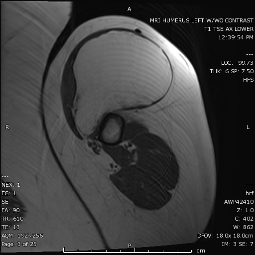
There is a large mass which follows fat signal intensity on this T1 non fat saturated axial MRI image of the left humerus. The masss is located within the biceps brachii muscle and is consistent with an intramuscular lipoma.
Contributed by Hassana Barazi, MD.