Discoid Lupus Erythematosus
- Article Author:
- Brianna McDaniel
- Article Author:
- Sukesh Sukumaran
- Article Editor:
- Laura Tanner
- Updated:
- 8/15/2020 11:38:27 PM
- For CME on this topic:
- Discoid Lupus Erythematosus CME
- PubMed Link:
- Discoid Lupus Erythematosus
Introduction
Lupus erythematosus is a multisystem disorder that predominately affects the skin. There are several types of cutaneous lupus. The most common types are acute cutaneous lupus (ACLE), subacute cutaneous lupus (SCLE), and discoid lupus (DLE). Dr. James Gilliam described the most commonly used classification of cutaneous lesions in lupus erythematosus. Gilliam segregated skin lesions into those that are specific and those that are not specifically based upon whether an interface dermatitis was seen on histopathologic examination. Within the category of specific cutaneous lesions, he subdivided these into acute cutaneous lupus erythematosus, subacute cutaneous lupus erythematosus, and chronic cutaneous erythematosus.
The most common subset of chronic cutaneous lupus erythematosus is DLE. These patients may or may not report photosensitivity, but lesions are frequently photo distributed and have a propensity to have secondary atrophy or scarring. Most patients with DLE do not have significant systemic disease. DLE can also occur as a manifestation of SLE in approximately 20% of patients. Other less common forms of chronic cutaneous lupus erythematosus include hypertrophic lupus erythematosus, tumid lupus erythematosus, lupus erythematosus panniculitis (LEP or lupus profundus), oral DLE, as well as DLE lesions on the palms and/or soles.[1][2][3]
Etiology
Lupus erythematosus is an inflammatory, connective-tissue disease of generalized autoimmunity characterized by pathogenic autoantibodies and immune complexes which are attributed to a loss of immune tolerance. For discoid lupus erythematosus without associated SLE (CDLE), the evidence does not show whether circulating inflammatory cells and autoantibodies are involved in the pathogenesis, but it is evident that the cutaneous inflammatory infiltrates are dominated by Th1, but not Th17, cells in contrast to systemic lupus erythematosus.[4][5]
Epidemiology
Lupus can occur in all age groups, but DLE occurs more frequently in women in their fourth and fifth decades of life. Twenty-five percent of patients with SLE may develop typical discoid lesions at some point during their illness, and 1% to 5% of patients with discoid lupus may develop SLE.
Ethnicity is also a major risk factor for developing LE, and its effect in some populations is almost as strong as that of gender. The prevalence of SLE is four-fold higher in African-American women as compared to Caucasian-American women (4 in 1000 versus 1 in 1000). In addition, African-Americans tend to develop the disease at an earlier age and have a higher mortality rate.[6][7]
Pathophysiology
The etiology of cutaneous lupus erythematosus is multifactorial with an interplay between genetic and environmental factors. Some contributing environmental factors include ultraviolet radiation (UVR), medications, cigarette smoking, and possibly, viruses. The interaction between these multiple factors triggers an inflammatory cascade of cytokine, chemokine, and inflammatory cell responses. Genes previously associated with SLE are TYK2, IRF5, and CTLA4 and also confer an increased risk for developing DLE.
An analysis of 405 patients by Bockle et al. found that smoking is highly associated with discoid lupus erythematosus. Bockle et al. hypothesized smoking may play a pathogenic role in cutaneous lupus erythematosus variants (DLE, tumid lupus) by the induction of apoptosis, its ability to stimulate T-cell proliferation, and increase photosensitivity. Another explanation might be that smoking provokes DNA damage, resulting in the formation of DNA adducts and the production of ds-DNA antibodies. Keratinocytes may also participate in lupus skin damage by increasing the apoptotic rate and the production of proinflammatory cytokines such as IFN-alpha and IL-6 for SLE, and IFN-lambda for DLE.[8][9]
Histopathology
The findings of histopathologic examination in cutaneous lupus vary based on the subtype. Overlap can be seen in the histologic findings among the various clinical phenotypes, particularly ACLE, SCLE, and discoid lesions. In cutaneous LE, basal cell damage (also referred to as vacuolar degeneration, hydropic change, or interface dermatitis) and lymphohistiocytic inflammatory infiltrates are commonly seen. In discoid lupus lesions, periadnexal inflammation, follicular plugging, and scarring are primarily seen in addition to the interface dermatitis.
Examination of the skin for deposits of immunoreactants is called direct immunofluorescence (DIF). DIF of lesional skin can be useful in establishing a diagnosis of cutaneous LE in cases where routine histopathology is equivocal. DIF does not replace routine histologic staining as the method of choice for establishing a diagnosis. In active lesions of DLE, DIF of lesional skin is positive in the majority of cases. The most characteristic DIF finding in cutaneous LE is antibody deposition at the dermal-epidermal junction and around hair follicles. These deposits are typically granular, and they are composed primarily of IgG and/or IgM.
The lupus band test (LBT) is a diagnostic procedure that is used to detect deposits of immunoglobulins and complement components along the dermo-epidermal junction in patients with lupus erythematosus (LE). LBT can be helpful in distinguishing systemic lupus erythematosus from chronic lupus erythematosus, because in SLE patients the LBT is frequently positive in both uninvolved and involved skin, whereas in CLE patients only the involved skin is positive. The LBT is positive on the lesional skin in 75% of patients. Ideal lesions for LBT for DLE are on the head and neck that have been present for at least a few months.
History and Physical
DLE is the most common form of chronic cutaneous erythematosus and can occur as localized form (80%) with lesions on the face, ears, and scalp or as disseminated DLE (20%) with lesions above and below the neck. The disseminated form of DLE, especially when involving the trunk, is associated with an increased risk of progression to SLE.
It is unusual for discoid lesions to be present below the neck without lesions also being present above the neck. Occasionally, discoid lesions develop on mucosal surfaces, including the lips, nasal mucosa, conjunctivae, and genital mucosa. Some patients with discoid lesions exhibit a photodistribution. Sun exposure seems to play a role in the development of lesions. However, patients can have discoid lesions on the sun-protected skin, and there is no clear association between sun exposure and their development.
The first morphological sign of DLE is a well-defined, annular erythematous patch or plaque of varying size followed by follicular hyperkeratosis, which is adherent to the skin. By removing the adherent scale, follicle-sized keratotic spikes similar to carpet tacks can be seen (“carpet tack sign”). The lesions slowly expand with active inflammation and hyperpigmentation at the periphery leaving depressed central atrophy and scarring, telangiectasia, and hypopigmentation. DLE can progress to irreversible scarring alopecia on the scalp. Although uncommon, a squamous cell carcinoma can develop in a longstanding discoid lesion.
Patients who present with discoid lesions may have associated arthralgias, but, over time, only approximately 10% to 20% of these patients eventually meet the classification criteria for SLE.
Evaluation
In the evaluation of DLE, the dermatologist should take a directed history, perform a cutaneous examination looking for signs of possible systemic disease. The diagnosis of DLE is made based on clinical features, but histology may be required to confirm the diagnosis.[10][11]
Autoantibodies to SSA/Ro, SSB/La, U1RNP, histones, and ssDNA, are common in patients with SLE, but they are not disease-specific. There are no other specific autoantibodies to differentiate the subtypes of CLE that are routinely used in practice. One further possible target of auto-antibodies is annexin 1, which has been suggested to play an important role in the prevention of autoimmune diseases. In a recent study, a significantly higher level of anti-annexin one antibodies was found in DLE patients, suggesting that anti-annexin one antibodies might be a new diagnostic marker for DLE.
Treatment / Management
Early treatment of discoid lupus lesions may lead to the total clearing of skin lesions, but treatment failure results in permanent scarring. Hair loss, depressed scars, and pigmentary changes are often disfiguring, particularly in darker-skinned people. Some general measures, such as sun avoidance and liberal application of sunscreen, are encouraged because cutaneous lesions are known to be exacerbated by sunlight. Smoking cessation is encouraged, as this can increase DLE disease activity. Studies demonstrate a statistically significant decrease in efficacy of antimalarial medication in individuals who have currently or ever smoked.[12][13][14]
Current first-line treatment for DLE consists of photoprotection in conjunction with topical or oral corticosteroids, topical calcineurin inhibitors, and systemic antimalarial therapy. Chronic DLE lesions that are not responsive to topical corticosteroids or topical calcineurin inhibitors may be responsive to intralesional corticosteroid injections. When DLE is refractory to these measures, other agents with varying degrees of proven efficacy are used. Currently, no medications have been approved specifically, and many of the drugs described in the literature were developed for use in other autoimmune disorders.
Antimalarials are immunotherapeutic and are considered first-line systemic therapy in CLE. Hydroxychloroquine (HCQ) and chloroquine with or without quinacrine are currently utilized in the treatment of DLE. HCQ is preferred over chloroquine due to the lower risk of side effects, specifically retinal toxicity.
Thalidomide, a potent teratogen, has been used in the treatment of DLE. An early report of its use in the treatment of DLE of 60 individuals treated with 50 to 100 mg per day found complete or marked regression in 54 individuals (90%) with disease relapse in 71% of individuals with medication discontinuation. Side effects included drowsiness, constipation, rash, edema, xerostomia, and 25% of individuals complained of slight to moderate polyneuritis symptoms. Lenalidomide is a thalidomide analog that may also prove useful in the treatment of DLE. Evidence suggests lenalidomide is effective in treating DLE and has a less severe side effect profile than thalidomide but may be similarly limited by a tendency to relapse once discontinued.
Other treatment modalities such as retinoids are vitamin A analogs with anti-keratinizing and anti-inflammatory effects are sometimes used in CLE, but documentation in the literature is limited. Immunosuppressive agents such as mycophenolate mofetil, azathioprine, intravenous immune globulin (IVIG), cyclophosphamide, and cyclosporine have all been trialed in the treatment of DLE but thought to be second-line when refractory to other treatments.
Differential Diagnosis
- Scleroderma
- Mixed connective tissue disease
- Cutaneous lymphoma
- Rheumatoid arthritis
Complications
- Arthritis
- Myositis
- Hypertension
- Renal failure
- Neuropsychiatric symptoms
- Pleuropericarditis
- Pancreatitis, mesenteric vasculitis
- Optic neuritis
Enhancing Healthcare Team Outcomes
Lupus is best managed by an interprofessional team of healthcare workers because the disorder can affect almost every organ in the body. Besides physicians, the role of the nurse, pharmacist, therapist, social worker, and a mental health counselor is vital. The key is to stress the importance of medication compliance. Patients should be educated about the importance of seeking help early when symptoms arise. At the first sign of renal dysfunction, medical help should be sought. Once renal failure is established, the only treatment is transplantation or dialysis. Patients should be told to avoid sunlight, stop smoking, eat healthily and remain active. Women of childbearing age should consult with an obstetrician before getting pregnant. Finally, joining a support group and/or following up with a mental health counselor is highly recommended.[15][16] (Level V)
Outcomes
Discoid lupus is an unpredictable and a highly variable disorder. While the condition is benign, it can cause devastating complications, which often lead to a high morbidity and a poor quality of life. The disorder frequently waxes and wanes. The outcome is much improved for patients with only skin and musculoskeletal involvement. The outcomes are worst for patients with CNS and renal involvement. Today, with treatment there is an 80% survival at ten years, but failure to comply with treatment can lead to early death. At some point in time, the majority of lupus patients will develop hypertension, lipid disorders, diabetes, infections, osteoporosis and malignancies like lymphomas and liver cancer. [17][18](Level V)
(Click Image to Enlarge)
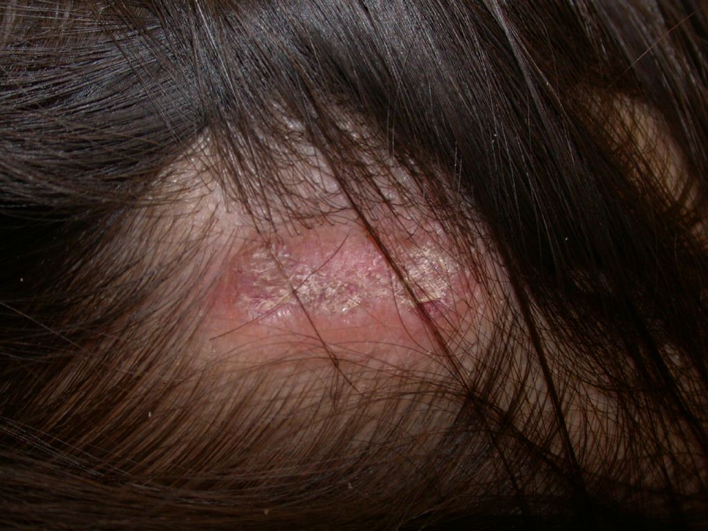
Discoid Lupus, Scalp
Contributed by DermNetNZ
(Click Image to Enlarge)
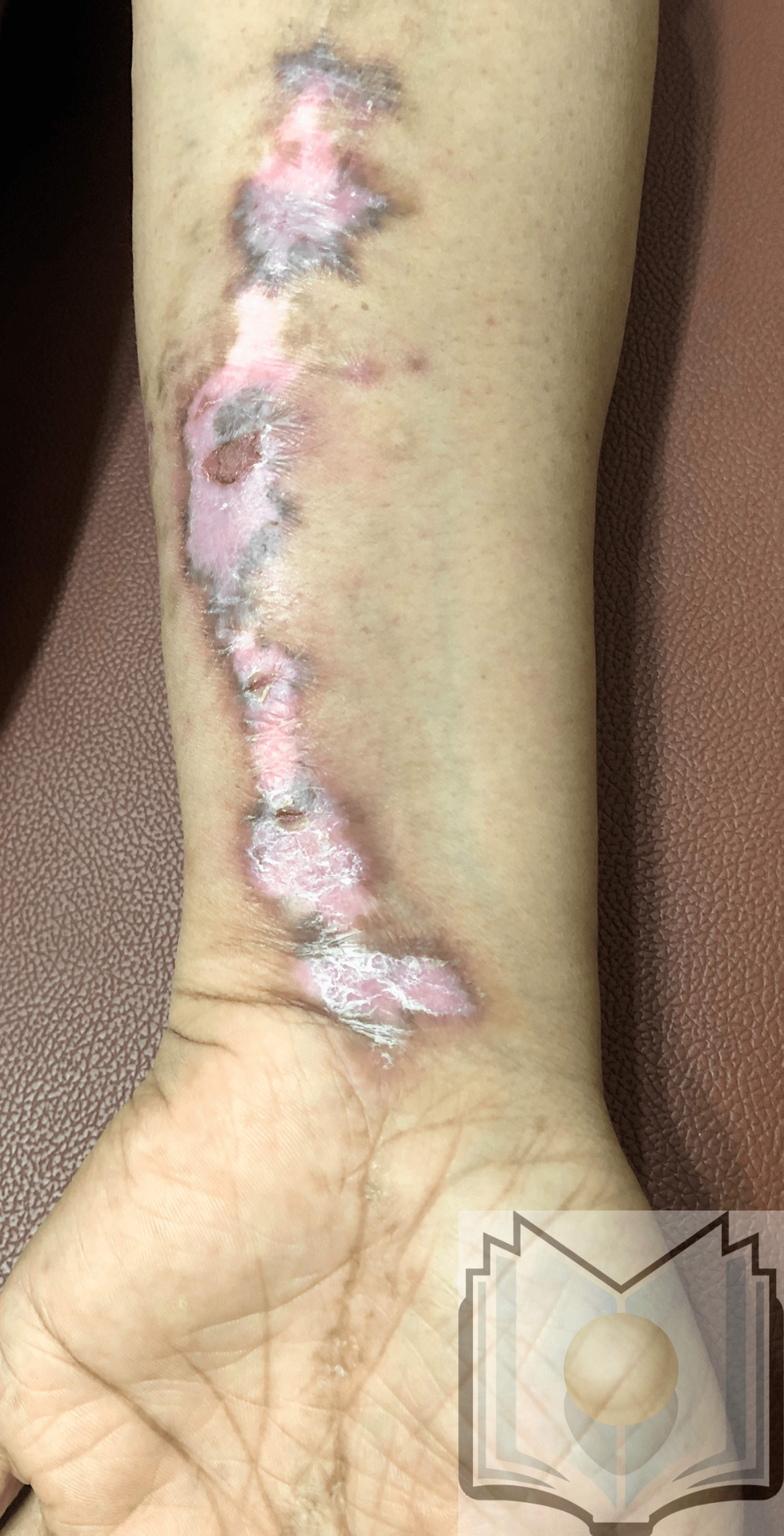
Linear Discoid Lupus Erythematosus
Contributed by Dr. Shyam Verma, MBBS, DVD, FRCP, FAAD, Vadodara, India