Malignant Otitis Externa
- Article Author:
- Mahmoud Al Aaraj
- Article Editor:
- Cecylia Kelley
- Updated:
- 8/10/2020 9:06:15 PM
- For CME on this topic:
- Malignant Otitis Externa CME
- PubMed Link:
- Malignant Otitis Externa
Introduction
Malignant otitis externa, although it is not a malignancy, it behaves and spreads like one, hence the name.[1]
The first case of MOE was reported in 1938, and the term ‘malignant otitis externa’ was described later by Chandler in 1968, due to its high fatality rate in that time.[2]
Malignant otitis externa is a life-threatening infection that mainly affects the external auditory canal and skull base, and the infection can also invade the stylomastoid and jugular foramina. Infection and inflammation can take different anatomical routes through the osteocartilaginous junction or osseous canal toward the mastoid process posteriorly, or toward the temporomandibular joint, parotid gland and cervicofacial spaces anteriorly, or medially into skull base.[3]
The disease ends up with osteomyelitis of the temporal bone after starting as simple otitis externa. Malignant otitis externa has an association with many serious complications, including cranial nerve involvement and increased rates of morbidity and mortality. Therefore, malignant otitis externa should be identified and treated urgently.[4][5]
Etiology
The most common causative organism is Pseudomonas aeruginosa.[6]
Fungi can also cause the disease by 5% to 20% of the general population, making it the second most common causative organism, with Aspergillus fumigatus being the most common cause of fungal MOE.[7][8]
Other organisms, such as Proteus mirabilis, Proteus sp., Klebsiella sp., and Staphylococci, have been isolated.[9]
Most of the patients diagnosed with MOE share varying degrees of immunosuppression, including patients with diabetes and immunosuppressed patients:
- Diabetes
- This is due to small-vessel vasculopathy and immune dysfunction associated with diabetes. Moreover, patients with diabetes have impaired response to antimicrobial agents, as their ear wax has a higher PH than normal people as well as lower lysozyme concentration. It has been reported that there is no difference in the prevalence of MOE between patients with type 1 or type 2 diabetes.
- Immunosuppressed patients (e.g., from HIV infection, chemotherapy, etc.).[10]
- Patients with AIDS have a different presentation, as they are usually younger, and Pseudomonas is not the most causative organism.
- Granulation tissue might not be found in the external auditory canal of patients with MOE and AIDS.
- These patients usually have a worse outcome than patients with diabetes.[11]
Therefore, do not exclude the diagnosis in young, non-diabetic, or immunocompetent patients.[9]
Epidemiology
The prevalence of malignant otitis externa has experienced a drop in recent years due to the development of the modern antimicrobials; however, the incidence of it did not reach the limit to be called as a rare disease.[1]
Malignant otitis externa has been reported in all age groups but is most common in patients who are older than 60 years.[12]
Males are more commonly affected than females.[13]
The infection had a higher incidence rate in areas with high humidity.[11]
Malignant otitis externa rarely occurs in pediatric patients.[14]
Pathophysiology
The infection can spread, causing bony erosions and invasion of distant tissue, using the fascial planes and venous sinuses, with the involvement of the skull base and the surrounding tissue leading to cranial nerve and intracranial structures invasion.
When the infection reaches the temporal bone through the fissure of Santorini, it invades the stylomastoid and the jugular foramina, containing the facial, glossopharyngeal, vagal, and accessory nerves. On the other hand, the spread through the osteocartilaginous junction to the subtemporal area causes invasion of the retrocondylar fat, the parapharyngeal fat, temporomandibular joint, and the masticator.[15]
The spread can be summarized as follows:
- Anterior: To masticator muscle, condylar bone marrow, parotid gland, facial nerve, temporal fossa, temporomandibular joint (TMJ), and the styloid foramen.
- Medial/crossed: To the parapharyngeal fat, nasopharyngeal muscle, glossopharyngeal, vagal and accessory nerves, sphenoid, clivus, jugular foramen, and petrous apex.
- Intracranial: When a dural enhancement is present in the intracranial compartment, the spread might reach sigmoid sinus, jugular vein, internal carotid artery, jugular fossa, and the dura.
- Posteriorly: To the mastoid process, however, there is no soft tissue invasion.[16]
Intravascular involvement can be seen as well, which is more common in fungal MOE; however, fungal MOE usually does not affect the temporal bone.[17]
Histopathology
Normally the external auditory canal (EAC) contains a layer of stratified squamous epithelium covering the underlying connective tissue along the osseous, while the osteocartilaginous junction and regions of the osseous canal are made of a thick subepithelial fibrous tissue. In patients with MOE, Biopsy of the external auditory canal (EAC) might show ulceration and loss of epithelium, with bacteria and inflammation reaching to the dense fibrous tissue. In the areas where the epithelium is not broken, reactive changes vary from mild hyperplasia to prominent pseudoepitheliomatous hyperplasia. Acute and chronic inflammation, including abscess formation, is common. In biopsy samples overlying the cartilaginous canal, the inflammation extends into the apopilosebaceous apparatus. Reactive capillary proliferation is evident within the granulation tissue. However, chronic granulomatous inflammation with granuloma formation is not associated with MOE. The identification of microorganisms is usually made by tissue staining. In case no organism has been identified, polymerase chain reaction (PCR) assay might help in detecting the causative organism.[3]
It can be difficult to differentiate very well-differentiated squamous carcinoma form MOE in the presence of pseudoepitheliomatous hyperplasia; therefore, the presence of MOE should not exclude malignancy, as these can occur concurrently.[18]
History and Physical
The best way to approach malignant otitis externa is to follow the diagnostic criteria which have been described by Cohen and Friedman, and it included major (obligatory) and minor (occasional) criteria as follows:
Major (Obligatory)
- Pain, which is often out of proportion to physical examination
- Edema
- Exudate
- Granulations, which can be seen in the external auditory canal
- Microabscess (when operated)
- Positive bone scan technetium-99 (99Tc)
- Failure of local treatment often more than 1 week
Minor (Occasional)
- Diabetes
- Cranial nerve involvement
- Positive radiograph
- Debilitating condition
- Older age
All of the major criteria must be present to diagnose malignant otitis externa; minor criteria alone are not sufficient to make the diagnosis.[19][20]
Cranial nerves examination should be done, as these are commonly affected.
- The facial (VII) nerve is most commonly affected
- The glossopharyngeal (IX) nerve
- The vagus (X) nerve
- The accessory (XI) nerve
- Rarely, the hypoglossal (XII) nerve[21]
Mental state examination should be performed, and if the mental state is affected, this indicates intracranial complications.[22]
Evaluation
Labs
- Blood
- White blood cell count can be normal or slightly elevated, and a left shift is not commonly present in MOE.
- Erythrocyte sedimentation rate and C-reactive protein
- Inflammatory markers are usually high in these patients and can be used as an indicator for the response for antimicrobial therapy. Once the disease has been identified, levels of ESR and CRP should be checked and followed regularly until they are back in the normal range. ESR starts to decrease within two weeks of starting treatment.[23]
- Blood glucose
- Blood glucose should be checked in patients with diabetes to assess their baseline, as MOE can affect the baseline glucose intolerance.
- Non-d patients should be evaluated for diabetes.[24]
- Culture and sensitivities from the external auditory canal
- Cultures from the ear drainage are to be done before initiating antimicrobial treatment.
- Tissue sampling has proven to be of high value in choosing the right antimicrobial treatment and in cases of recurrent otitis externa.
- Biopsy of the external auditory canal should be obtained to exclude other causes, such as malignancy or cholesteatoma.[23]
Imaging
- Computerized tomography
- The advantage of CT scanning is in detecting bony erosions and demineralization.
- CT findings usually include obliteration of the fat planes in the subtemporal area and destruction of the bony cortex of the mastoid.[25]
- Magnetic resonance imaging
- MRI is superior to CT with detecting anatomical locations and invasion of soft-tissue components.
- MRI is also better in evaluating intracranial complications such as thrombosis and intracranial spread.
- However, it is hard to differentiate between active inflammation and resolving infection; therefore, many studies suggested that the use of CT and MRI imaging does not correlate to the prognosis and outcome of the disease.
- Gallium citrate Ga 67 scan
- Ga67 is a useful tool to monitor the resolution of the disease. The affected area usually shows increased uptake. However, the lesion to non-lesion ratio should be calculated to have more accurate results regarding the activity of malignant otitis externa.[26]
- Technetium Tc 99 methylene diphosphate bone scanning
- Tc99 is useful for the initial evaluation of the disease, but the test is not useful in assessing the prognosis of the disease, as it stays positive for a long period, even after the resolution of the infection.
Nuclear studies are not sufficient to show the anatomical extent of the disease. CT and MRI should be accompanied by SPECT bone imaging for the initial diagnosis.
SPECT, in addition to gallium 67 scan, is the investigation of choice to assess the progress of the disease.[27]
Treatment / Management
Antibiotic Therapy
- The treatment depends on the administration of systemic anti-microbial therapy with anti-pseudomonal activity, typically a fluoroquinolone in cases caused by pseudomonas aeruginosa, however, with the widespread use of ciprofloxacin in upper respiratory tract infections, many resistant cases have been reported in the literature.[17] Oral ciprofloxacin can be used in the outpatient setting in cases which are not complicated by cranial nerve involvement, or the need for admission for pain control and diabetes. Resistant cases can be treated with parenteral antibiotics using antipseudomonal beta-lactam antibiotics with or without an aminoglycoside.
- Although most cases are caused by Pseudomonas aeruginosa, other organisms have been identified as a cause of MOE, and therefore the antibiotic management should be according to the isolated organism.
- The duration of treatment depends on the response to therapy, which is evaluated using a gallium citrate Ga 67 scan every 4 to 6 weeks during treatment.
- The treatment should be stopped one week after a normal gallium citrate Ga 67 scan.
Hyperbaric Oxygen Therapy
- Although it has been widely used in the treatment of MOE, many studies showed no added benefit for using it as an adjuvant for medical or/and surgical therapy.[28][29]
Surgical Therapy
- Surgical management is reserved for the patients in whom medical therapy has failed to cure the disease. Surgical therapy includes local debridement, removal of bony sequestrum, and abscess drainage. However, there is no indication for facial nerve decompression in patients with facial nerve involvement; finally, the surgery did not show any prognostic benefits.[30][31]
Differential Diagnosis
Malignant otitis externa can be mistaken by many other conditions, these include:
- Localized otitis externa
- Acute diffuse otitis externa
- Chronic otitis externa
- Carcinoma of the ear canal
- Aspergillus skull-base osteomyelitis[24]
Prognosis
The duration of diabetes, the presence of ESR and CRP, and imaging studies results have all affected the prognosis of MOE.[31]
The poorer prognosis was seen in patients who experienced any of the following:
- Facial nerve involvement
- Additional cranial nerve involvement
- Non-cranial nerve neurological involvement
- Extensive granulations (or edema) in the external auditory canal
- Bilateral symptoms
- Aspergillus species as the causative organism[32]
Elderly patients had a higher chance of developing complications and had a higher mortality rate compared to patients from younger age groups.[33]
Recurrence
The recurrence rate of MOE is high, reaching 15% to 20% when ESR starts to rise again.[22]
MOE can recur up to one year after treatment; therefore, the patient should be followed up regularly for one year before being considered cured.[14]
Mortality
With the advanced treatment modalities, the mortality rate dropped from a high of 50% in the past to 10% to 20%.[34]
Complications
Complications of MOE can occur due to the invasion of the surrounding structures and cranial nerve involvement. Most commonly, the facial nerve is affected; however, glossopharyngeal, vagus, accessory, or hypoglossal nerve can also be involved.[21]
Skull base osteomyelitis arises when the disease spreads to reach the sphenoid, occiput, and clivus, rather than being confined to the temporal bone.[15]
Intracranial involvement might also happen to range from confusional state to severe complications such as meningitis, thrombosis, or death.[22]
Consultations
Deterrence and Patient Education
Patients should be aware of the consequences of the disease and how to control the factors that might increase the risk of having malignant otitis externa. There are modifiable and non-modifiable risk factors for the disease, studies have shown that diabetes, as a modifiable risk factor, can affect the progress of the disease. Therefore, patients with diabetes should have tight glycemic control to avoid getting the infection.[31]
Moreover, patients should avoid the causes of otitis externa, like swimmer’s ear, which might lead to malignant otitis externa.[5]
Enhancing Healthcare Team Outcomes
Malignant otitis externa is a serious infection and can lead to many severe complications, therefore, a team approach must be carried while taking care of these patients in order to improve the outcome for the patients and decrease the rate of complications. A team of an otolaryngologist, radiologist, infectious disease specialist, microbiologist, primary care, and an endocrinologist should be involved looking after patients with MOE. All working together can enhance better outcomes and work on the comorbidities that might bring up the disease.[27][17][31][6][9]
(Click Image to Enlarge)
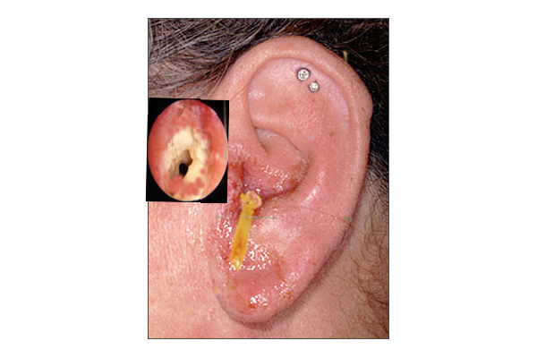
otitis externa
image courtesy S bhimji MD