Meckel Diverticulum
- Article Author:
- Jason An
- Article Editor:
- Christopher Zabbo
- Updated:
- 8/11/2020 8:18:52 PM
- For CME on this topic:
- Meckel Diverticulum CME
- PubMed Link:
- Meckel Diverticulum
Introduction
Meckel diverticulum is caused by the incomplete obliteration of the omphalomesenteric duct in the developing embryo. It is the most common congenital anomaly of the gastrointestinal (GI) tract.[1] The incomplete obliteration of the duct results in a diverticulum in the small intestine. Often, these are completely asymptomatic.[2] Acid secretion from the ectopic gastric mucosa within the diverticulum can result in GI bleeding and abdominal pain. It is useful to remember the rule of 2s.
Meckel diverticulum occurs in 2% of the population, 2% are symptomatic, children are usually less than 2 years, affects males twice as often as females, is located 2 feet proximal to the ileocecal valve, is 2 inches long or less, and can have 2 types of the mucosal lining.
Etiology
Meckel diverticulum is caused by the incomplete obliteration of the omphalomesenteric duct which connects the yolk sac to the gut in the developing embryo. It provides nutrition until the placenta forms. At about 7 weeks of gestation, the duct separates from the intestine. If the duct fails to partially or entirely separate and involute, it can result in an omphalomesenteric cyst, omphalomesenteric fistula that drains through the umbilicus, or a fibrous band from the diverticulum to the umbilicus which can cause an obstruction.[3] If there is no additional attachment, it forms into a Meckel diverticulum. The “rule of 2s” has been used to describe a Meckel diverticulum conveniently. It is the most common congenital GI anomaly which occurs in about 2% of infants.[1] It usually measures 2 inches long and is located in the ileum approximately 2 feet from the ileocecal valve. It is twice as common in females. It can contain 2 types of tissue (gastric or pancreatic). It is a true diverticulum because it contains all the layers of the small bowel wall. The diverticulum can sometimes have ectopic tissue within the walls. The embryonic origin of the ectopic tissue is unknown. It is estimated approximately 15% of patients will have ectopic tissue within the diverticulum.[1]
Epidemiology
Meckel diverticulum is the most common congenital anomaly of the GI tract. There is no clear familial predisposition. Patients with other malformation of the GI tract, nervous system, or cardiovascular system have increased the risk of having a Meckel diverticulum. The prevalence of Meckel diverticulum in the general population is estimated to be approximately 2%; however, it is difficult to determine the exact number because many patients are asymptomatic.[1] The patient often becomes symptomatic in the first decade of life with an average age of 2.5.[2]
Pathophysiology
Many patients with Meckel diverticulum are asymptomatic. Risk factors for increased risk of developing symptoms include age younger than 50, male gender, diverticulum greater than 2 cm in length, the presence of ectopic tissue, broad-based diverticulum, and fibrous bands attached to the diverticulum.[2] The pancreatic bicarbonate in the duodenum typically neutralizes the acid secreted by normal gastric mucosa. In a Meckel diverticulum, the ectopic gastric mucosa secretes an acid that is not neutralized and resulting in ulceration of the adjacent mucosa leading to painless rectal bleeding. The ectopic mucosa can also originate from the pancreas, jejunum, or a combination of mucosa.[4] The site of the bleeding is usually distal to the diverticulum and not within the diverticulum. The presence of a fibrous band attached to the diverticulum can result in small bowel obstruction. The diverticulum can also act as a lead point for an intussusception leading to small bowel obstruction.[5] The incarceration of the Meckel diverticulum can also result in a small bowel obstruction. Inflammation in the diverticulum can result in Meckel diverticulitis with perforation.
History and Physical
Many patients who have a Meckel diverticulum will never find out they have it. It is often found incidentally on imaging studies. If patients do develop symptoms, they usually present in the first 10 years of life with an average age of 2.5 years with painless rectal bleeding.[2] The rectal bleeding is typically described as currant jelly or the color of brick. Children typically present with the classic “currant jelly” colored stool while adults typically present with melena.[6] The bleeding usually resolves without intervention. As the patient becomes hypovolemic, the splanchnic vessels vasoconstrict to prevent further bleeding. Patients can present with a variety of GI symptoms other than painless rectal bleeding. It should be suspected in children with recurrent or atypical intussusception, a patient with symptoms of appendicitis after their appendix has been removed, and adults with an unclear source of GI bleeding.[1] Although it is classically described as painless rectal bleeding, some patients may present with abdominal pain.
Evaluation
Clinically, Meckel diverticulum should be suspected in any child younger than 2 years of age with painless rectal bleeding. It accounts for approximately 50% of all lower GI bleeding in children younger than 2 years of age.[6] Plain abdominal x-ray is of very low yield. Even barium studies rarely fill the diverticulum. The most sensitive test is a Meckel radionuclide scan (commonly known as Meckel scan).[7] It is a nuclear study done by injecting technetium-99m which is absorbed by the ectopic gastric mucosa allowing for visualization of the Meckel diverticulum. The uptake of the dye can be enhanced using cimetidine, ranitidine, or glucagon. In patients with active bleeding, a tagged red blood cell (RBC) scan can also be used to detect a Meckel diverticulum. A CT scan of the abdomen and pelvis may show evidence of inflammation or obstruction at the diverticulum. More invasive methods include mesenteric angiography as well as exploratory laparoscopy.[8]
Treatment / Management
If the patient has had significant blood loss, the patient should undergo volume resuscitation. Patients may require a blood transfusion if a significant amount of blood is lost. The treatment of symptomatic Meckel diverticulum is surgical excision. The diverticulum can be removed via laparoscopic or open technique.[9] The diverticulum should be excised along with the adjacent ileum. There is a growing trend to perform the excision laparoscopically rather than using the open technique.[9] The treatment of asymptomatic Meckel diverticulum is controversial (surgery versus observation). When a Meckel diverticulum is incidentally discovered during abdominal surgery for another condition, most surgeons recommend removal.[9]
Differential Diagnosis
The differential diagnosis is broad in can including any cause of GI bleeding. Massive GI bleeding is uncommon in childhood. Stool may be mistaken for hematochezia if children ingest bismuth, iron, or spinach. A hemoccult test will show that the stool will be negative. In infants, swallowed maternal blood from bleeding nipples, milk protein allergy, intussusception, and anal fissures can commonly cause rectal bleeding. Necrotizing enterocolitis should be on the differential in neonates and premature infants. In older children, other common causes of rectal bleeding include colitis, gastroenteritis, HSP, HUS, intussusception, inflammatory bowel disease, and vascular malformation.
Complications
The most common complication of Meckel diverticulum in children is rectal bleeding resulting in anemia.[5] The most common complication in adults is a small bowel obstruction.[10] The etiology of the obstruction may be secondary to the omphalomesenteric band, internal hernia, volvulus around the vitelline duct remnants, and intussusception in which the diverticulum acts as the lead point.[5] The diverticulum can become inflamed resulting in Meckel diverticulitis with perforation and peritonitis.
Enhancing Healthcare Team Outcomes
To provide the best care of a patient with Meckel diverticulum, a team approach should be used. There are a number of potential complications and the nurses should be educated by the physicians to watch for them and encouraged to report any abnormal findings. In particular, these patients are at risk of developing a perforation and acute peritonitis. [Level V]
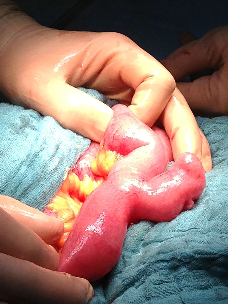
(Click Image to Enlarge)

The picture shows a Meckel Diverticulum (white arrow).
Contributed by Bruno Bordoni, PhD.
(Click Image to Enlarge)
Meckel diverticulum
By Milliways - Own work, CC BY-SA 3.0, https://commons.wikimedia.org/w/index.php?curid=23309979