Nasal Fracture
- Article Author:
- Charles Goodmaker
- Article Editor:
- Orlando De Jesus
- Updated:
- 8/10/2020 10:52:43 PM
- For CME on this topic:
- Nasal Fracture CME
- PubMed Link:
- Nasal Fracture
Introduction
Fractures of the naso-orbital-ethmoid (NOE) complex involve the bones that form the NOE confluence, which includes the anterior cranial fossa, frontal bone, bones of the ethmoid and frontal sinuses, nasal bones, and orbits.[1] They often occur alongside injuries to other parts of the face and body but can occur in isolation. Road traffic accidents and physical violence are the leading causes of these injuries, but this picture is changing with improved vehicle and road safety.[2]
Knowledge of regional anatomy is fundamental in understanding assessment and management. The approach to these injuries starts with the advanced trauma life support approach, as these patients can have injuries to critical structures such as the airway. Further assessment relies on thorough clinical assessment aided by radiological imaging. The operative intervention depends on the classification of the NOE complex fracture, which is based on the status of the medial canthal tendon.[3] Meticulous primary surgical correction is key in restoring aesthetic features and preventing future complications of trauma. Operative approach and exposure is carefully considered to balance the need to correct the deformities but also to prevent further aesthetic disruption and complications.[4]
Etiology
The NOE fracture pattern is caused by forceful, direct trauma to the central midface. Due to the high energy involved, these fractures often occur in combination with injuries to other parts of the face and body. Road traffic collision is the most common cause of NOE fractures, especially with motorcycles.[5]. Fortunately, it appears to have reduced in frequency. The introduction of seatbelts and airbags has helped to decrease the incidence of facial fractures.[2] Physical assault, sport, and horse kicks have also been associated with this fracture pattern.
Epidemiology
NOE fractures make up 5% of all facial fractures in adults, with young males most commonly affected.[6][7] In the pediatric population, a higher 16% incidence exists.[8] Facial fractures overall in the United States are thought to cost around 1.06 billion dollars, with 93,808 associated hospital days.[9]
Pathophysiology
The naso-orbital-ethmoid complex is a confluence of structures made up of the nasal bones, nasal process of the frontal bone, frontal process of the maxilla, lacrimal bone, lamina papyracea of the ethmoid bone, and sphenoid bone.[1] The medial canthal tendon (MCT), also called medial palpebral tendon, is a band of fibrous tissue originating from the medial palpebral part of the orbicularis oculi muscle as well as the superior and inferior tarsus of the eyelids. The MCT splits and surrounds the lacrimal fossa, which includes the lacrimal duct before inserting into the lacrimal crest of the maxilla anteriorly and the lacrimal bone posteriorly.[10] The facial skeleton is formed of four paired vertical and four horizontal buttresses, which are columns of bone that provide structure to surrounding tissues and help provide a form to the face. The NOE complex incorporates the medial vertical and inferior and superior transverse buttresses. These become the focus during surgical fixation. It has been suggested that NOE fractures are the result of the facial bone design. The nasal bones transmit a force posteriorly to thinner bones, which crumple to transmit the force to the ethmoid sinuses, thus sparing the contents of the cranial vault and orbital contents.[11] However, there is disagreement on whether facial bone fractures provide any protection to the brain itself.[12]
Sensory innervation to the NOE region is by the ophthalmic and maxillary branches of the trigeminal nerve. Motor innervation is by the zygomatic branches of the facial nerve. Autonomic supply consisting of sympathetic and parasympathetic fibers innervate both intraocular and external structures. The lacrimal nerve transmits both fiber types to the lacrimal gland. Sympathetic fibers also provide innervation to the smooth muscle of the lids widening the palpebral fissure when stimulated.
Arterial supply to the region originates from the internal and external carotid arteries. The facial and maxillary arteries are given off by the external carotid artery and supply the majority of the face. The ophthalmic artery originating from the internal carotid supplies the orbital structures. The ophthalmic artery also gives rise to the anterior and posterior ethmoidal arteries which supply the ethmoid sinus and terminate in the nasal cavity where they anastomose with the sphenopalatine artery to form Little’s area which is prone to bleeding. Venous drainage follows a similar pattern to the arterial supply. The venous blood drains into the cavernous sinus, which can produce complications from thrombosis secondary to trauma or infection.[13]
History and Physical
An advanced trauma life support approach should be adopted when assessing patients with suspected facial trauma. Airway patency and stability are a key priority alongside cervical spine immobilization and hemorrhage control, as 10% of complicated facial fractures are associated with significant bleeding.[14] Full neurological and ophthalmic examinations are indicated as two-thirds of facial fractures are associated with some form of ocular injury.[15]
Examination of the NOE complex begins with a visual and manual inspection, which often will reveal severe swelling and periorbital ecchymosis, which sometimes make examination challenge. Excessive tear overflow in the eye and face (epiphora) can be associated with lacrimal duct damage or obstruction. A more reliable assessment of lacrimal duct function is using dye tests or dacryocystography (radiological contrast assessment of the lacrimal apparatus) if epiphora is persistent following surgery.[16] Nasal bone fracture and depression can result in a decreased nasal dorsal projection with an associated upturn of the nasal tip.[17] Epistaxis with associated mucosal nasal tearing may be present with or without cerebral spinal fluid (CSF) rhinorrhoea, as a result of anterior cranial fossa fracture. CSF presence can be confirmed with a beta-transferrin test, which is more accurate than the halo sign (CSF fluid on filter paper forming a halo pattern). Nasal patency can be crudely assessed with a metal object placed under each nostril.
Assessment of the MCT is a fundamental aspect of discerning the severity of an NOE complex injury. Telecanthus (increased distance between to the two medical canthi) with equal interpupillary distance is a sign of MCT rupture. Intercanthal distance is on average 30 to 31 mm, and the interpupillary distance is 60 to 61 mm. An intercanthal distance greater than 40 mm is noticeable, and an indication for surgical correction.[18] The bow-string test involves palpating the nasal root whilst retracting the eyelid inferior-laterally, the eyelid will have greater laxity, and a fractured segment may be palpable if the MCT is compromised.
Evaluation
Investigations
The examination may be initially limited by severe pain and gross swelling of facial structures. Computed tomography (CT) scan of the head provides definitive details of soft tissue and bony injuries. The use of both 2D and 3D images on coronal and axial views aids the diagnosis and staging of NOE complex fractures and assists the operating team in planning corrective operations.[19] The approach, degree of exposure, and equipment required are highly dependent on the CT scan. The study also outlines injuries of other structures in the head. In a trauma setting, it is likely to be performed early once the patient is stabilized with an advanced trauma life support approach.
Classification
The Markowitz and Manson system is the most widely used classification system of NOE complex fractures and has replaced other systems by making the integrity of the MCT a key feature of fracture severity.[3] This classification system relies on both CT scan and clinical examination which outline the status of NOE complex bony structures and MCT integrity, respectively:
- Type 1: The MCT is attached to a central fracture segment.
- Type 2: The MCT is attached to a comminuted central fracture segment.
- Type 3: The MCT is detached from a comminuted central fracture segment.
Treatment / Management
NOE fractures require surgical fixation and/or reduction to restore the aesthetic features of the face. If the medial canthal tendon is avulsed, then this will need to be reduced. The approach will depend on the severity and distribution of fractures, and an effort is made to use the smallest incision to provide the greatest exposure of tissue and bone.[1] The consideration of the incision site is very important as it can have a great aesthetic impact. Sometimes facial lacerations or pre-existing scars can be utilized. Pre-injury photographs can be of assistance to surgeons in planning reduction and fixation as so to return, as close as possible, the face back to its original form.
The coronal incision provides good exposure of the mid-upper face and is the gold standard approach to NOE fractures involving the frontal sinus. The midface degloving approach provides greater exposure of the midface. This technique, however, is associated with a number of complications, such as anesthesia and nasal deformity. Reduction and fixation of bony segments are sought before soft tissues are corrected.[20] In type 1 NOE fractures, closed reduction can be achieved. Type 2 and type 3 fractures will require exposure of the fractured segments with open reduction and internal fixation. A titanium mesh is used to stabilize the medial orbital wall, however, absorbable meshes can be used.[21] Microplates and screws are used to fix and stabilize bones and ensure the stabilization of the horizontal and vertical buttresses. Frontal sinus involvement requires additional repair of fractured walls and repair of open meninges if present. The sinuses are obliterated to avoid future mucocele formation with a variety of techniques. Pedicled flaps, autologous grafts (adipose tissue, bones, and muscles), xenografts, and biomaterial can be used for this purpose. Alternatively, in more severe sinus injuries, cranialization can be utilized - this is the removal of the posterior frontal sinus wall allowing brian tissue to fill the space.[22]
In type 3 NOE complex fractures reduction of the MCT (canthopexy) is achieved by transnasal wiring, which is performed by drilling a small hole into the medial orbital wall and tethering the MCT with a wire. The use of a needle to secure the MCT in lieu of a drill is sometimes required for unstable comminuted fractures of the medial orbital wall.[23]
Disruption to the lacrimal pathways is a common complication of midfacial trauma, with epiphora reported in nearly half of cases immediately postoperatively. However, permanent epiphora is relatively uncommon.[24] As such, secondary correction with dacryocystorhinostomy is preferred six months after fracture fixation.[16] The aim is to correct tear drainage and prevent future mucocele formation.
Differential Diagnosis
It should be noted that facial fractures seldom occur in isolation of injury to other parts of the face or body. Other groups of facial fractures can occur in isolation or alongside NOE complex fractures:
- Zygomaticomaxillary complex fractures may occur in association or independently. They are associated with a lateral forceful blow to the mid-lower face.
- LeFort fractures are defined as the separation of the midface from the skull base and graded from type 1-3. They must involve the pterygoid plates, and sphenoid bone as these connect the skull base to the midface.
- Orbital floor fractures, known as a blowout fracture, in which the fracture extends out from the globe towards the palate. Extraocular tissue/muscle herniates through the defect, causing diplopia and ophthalmoplegia.
- Panfacial fractures, which involve all three upper, middle, and lower regions of the face.
Prognosis
The majority of patients who are treated with primary corrective surgery have a positive aesthetic result. In severe type 3 fractures, some patients experience continued nasal deformity.[25] It is well documented that failure to correct complications, such as telecanthus on the first attempt, proves very challenging for a repair in the future.[20]
Complications
- Airway compromise and hemorrhage.[14]
- Nasofrontal duct and or lacrimal duct disruption as a result of direct damage or due to displaced fracture segments.[16]
- Facial deformity, as full correction of telecanthus or nasal depression can be difficult to achieve, and some patients will retain a degree of asymmetry. Depending on the surgical approach, patients may experience temporary or permanent paralysis and or anesthesia of the forehead. Scars that cannot be hidden in the hairy scalp or skin folds may be prominent.[26]
- Infection of the incision site, soft tissues, and meninges are recognized complications from these injuries.[26]
- Mucocele formation is a complication of sinus or lacrimal drainage disruption and can become infected.[26]
- Mental health, as patients with facial injuries are at greater risk of developing post-traumatic stress disorder or anxiety-related disorders. Particularly those who were victims of assault.[27]
Postoperative and Rehabilitation Care
Following corrective surgery, patients should be informed not to blow their nose for at least 10 days following NOE fractures.
Heavy lifting, straining, and vigorous exercise should be avoided postoperatively.
Eye drops are provided to maintain eye hydration and stabilize the tear film layer.[1]
Control of postoperative pain and associated nausea is encouraged.
A soft tissue bolster is placed over the nasal root covering the medial canthus to ensure healing of the soft tissue contour, which can be affected if a serous fluid collection develops.[28]
Perioperative antibiotic therapy is dependent on the type of injury, mechanism, and operating team. Contaminated wounds, exposure of the meninges, and/or evidence of CSF leakage are indications for prophylactic antibiotics to prevent wound infection and bacterial meningitis, respectively.
Monitoring of airway, neurological status, and ophthalmological sequelae take precedence.
Postoperative imaging may be indicated to ensure proper correction has been achieved.
Maintenance of fluids may be required until the patient is able to swallow safely, and nutrition optimized with supplemental drinks.[28]
Follow up to check wound healing, the progress of soft tissue correction, and the patient's functional status is mandatory.
Deterrence and Patient Education
Advances in motor vehicle safety have reduced the rate and severity of facial injuries observed in developed countries, with the universal introduction of deployable airbags playing a vital role. Continued education should be directed at patient populations most likely to be affected by severe facial injuries, as this may further reduce the rate and severity of facial injuries. Improving young driver awareness and teaching about the often underappreciated physical implications of violence would impact the key demographic population affected by these injuries.
Enhancing Healthcare Team Outcomes
Naso-orbital-ethmoid fractures are complex. Health and functional status are severely affected by facial fractures.[29] [Level 3] As such, they require input from a vast array of interprofessional team members to ensure the patient receives the highest level of care. This team includes the emergency group to stabilize a patient following trauma, maxillofacial surgeons, orthopedic surgeons, neurosurgeons, radiologists who performed detailed imaging, anesthesiologists, dieticians, and psychologists. It has been shown that timely primary surgical intervention can improve patient outcomes.[25] [Level 5] Such intervention can only be achieved if all members work in an efficient and synchronized fashion. The care given in the postoperative period is vital in improving functional impact.
(Click Image to Enlarge)
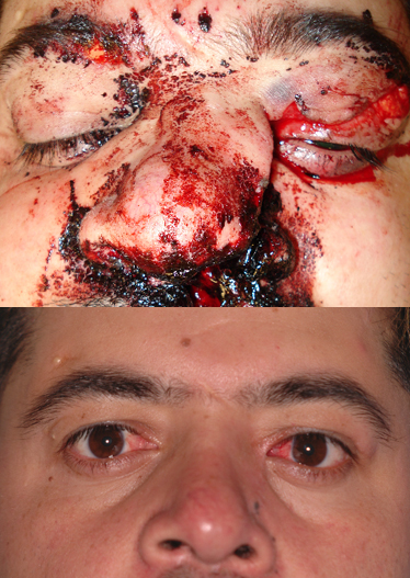
A 32-year old male had naso-orbito-ethmoid (NOE) fracture repair 12 months ago, after a motor vehicle accident. He had open repair of his nasal fractures and transnasal wires placed. He now presents with a complaint of intermittent tearing from the right side since the surgery and a mucoid discharge from the left side. Photos show appearance at time of injury (above) and appearance 12 months after repair.
Contributed by Prof. Bhupendra C. K. Patel MD, FRCS