Nasotracheal Intubation
- Article Author:
- Thomas Folino
- Article Author:
- George Mckean
- Article Editor:
- Lance Parks
- Updated:
- 8/13/2020 7:26:33 PM
- For CME on this topic:
- Nasotracheal Intubation CME
- PubMed Link:
- Nasotracheal Intubation
Introduction
Nasotracheal intubation (NTI) involves passing an endotracheal tube through the naris into the nasopharynx and the trachea; most commonly after induction of general anesthesia in the operating room. The use of NTI permits administration of anesthetic gases without obfuscation of intraoral anatomy and is commonly used for procedures including dental, oropharyngeal and maxillofacial operations. [1][2][3]NTI is an essential skill for anesthesia providers. Due to the potential complications in performing NTI, it is recommended that NTI not be attempted by anyone who is not skilled at orotracheal intubation as well.[4][5]
Anatomy and Physiology
Being able to perform NTI properly requires knowledge of the anatomy of the nasal vestibule, nasopharynx, oropharynx, and hypopharynx.
The nasal cavity begins at the anterior nares and ends at the posterior end of the nasal septum where it channels into the nasopharynx via the posterior nasal apertures (choanae). The nasal cavity sits above the oral cavity and hard palate and rests below the skull base.
The hard palate provides the base of the cavity that runs horizontally and directly behind the anterior nares. The ceiling of the nasal cavity is formed from the narrow cribriform plate of the ethmoid bone. Lastly, both left and right lateral walls are established by the medial wall of the respected orbit superiorly and the maxillary sinus inferiorly.
The lateral nasal walls incorporate three structures, the turbinates, which project into the nasal passages as ridges of tissue and are responsible for maintaining moisture and warmth in the nasal cavity as air flows through.
The inferior turbinate is the largest of the 3 and projects along the complete lateral nasal wall. The inferior turbinates are often responsible for blocking nasal airflow when they are enlarged or inflamed. The middle turbinate projects into the central nasal cavity adjacent to the nasal septum. Finally, the superior turbinate, the smallest of the three, attaches to the skull base superiorly and the nasal wall laterally.
The nasal cavity is separated by a nasal septum consisting of both a cartilaginous part that sits anteriorly and a bony portion that rests more posteriorly. This septum separates the anterior nasal pathway into a left and a right side, where these 2 cavities eventually coalesce to form a single continuous cavity in the back of the nose (the nasopharynx).
The nasal cavity is lined by respiratory mucosa (histologically described as ciliated pseudostratified columnar epithelium) lying on an extremely vascular stroma. These cells produce serous secretions that aid in humidification of inspired air. The cilia help to trap unwanted debris from entering the lungs.
Due to the high vascularity of the nasal cavity, minor trauma to any part of the tissue can cause bleeding to occur (epistaxis). The anterior nasal septum is particularly susceptible to developing epistaxis owing to the superficial location of the arterial plexus. This plexus is known as Kiesselbach’s area and is supplied by branches of the anterior and posterior ethmoid, superior labial, sphenopalatine and greater palatine arteries.
While considering normal nasal cavity anatomy, it is also important to understand that anomalies do often exist. Septal deviation is perhaps the most common anomaly which usually involves the cartilaginous aspect of the nasal septum. The deviation is most often due to trauma but can also be caused by continuous nasal congestion from recurrent sinus infections. Other variations to normal anatomy include conditions that result in unilateral obstruction such as nasal polyps, concha bullosa, and spurs. It is important to consider these anomalies during pre-anesthetic evaluation to minimize any complications as most of these variations will result in changes to airflow dynamics inside the nasal cavity. Nasal polyps or spurs may be unilateral which may dictate which side of the nose is more amenable to NTI.[6][7][8]
Indications
Indications for NTI include, but are not limited to the following:
- Intraoral and oropharyngeal surgery
- Complex intra-oral procedures involving mandibular reconstructive procedures
- Rigid laryngoscopy
- Dental surgery
- Maxillofacial or orthognathic surgery
Contraindications
Absolute contraindications include:
- Suspected epiglottis
- Midface instability
- Previous history of old or recent skull base fractures
- Any known bleeding disorder that could predispose the patient to severe epistaxis
- Choanal atresia
- Patients that have experienced high-speed trauma or isolated facial trauma may have undiagnosed skull fractures that may result in nasotracheal tube placement into the brain. It is best to avoid NTI in these patients.
Relative contraindications include:
- Anything that could compromise the nasal air passage (large nasal polyps, foreign bodies)
- Recent nasal surgery
- History of frequent episodes of epistaxis
Equipment
Some of the necessary equipment needed to perform a nasotracheal intubation includes the following:
- Endotracheal tube (Nasal RAE or standard endotracheal tube)
- Lidocaine jelly or a water-soluble lubricant
- Magill forceps
- Laryngoscope
- Vasoconstricting nasal spray (oxymetazoline 0.05% or phenylephrine nose drops 0.25% to 1%)
- Syringe to inflate cuff
Preparation
A pre-anesthetic evaluation must be performed for each patient undergoing a procedure requiring anesthesia with a particular focus on identifying any potential risks or complications related to the upcoming procedure and composing an individualized plan for patient care. Often, the patient can relay important information regarding unilateral restriction or congestion and give some direction as to which side should be used for the NTI. If the patient interview does not yield information related to relative patency of one side versus the other, then either side of the nose may be used.
An anterior rhinoscopy may be performed (this is not a common practice) which gives the provider the ability to visualize the anterior portion of each nasal cavity. The main limitation of anterior rhinoscopy is the inability to provide any information regarding the posterior nasal cavity. To fully assess the pathway, a flexible fiber-optic bronchoscope may be passed into the nasopharynx.[9][10]
Technique
Once the choice of laterality is determined, the provider may proceed with NTI. The first step to performing an NTI is a generous application of vasoconstricting spray bilaterally. Anesthetic topicalization may be applied via spray or by use of a lubricant that is impregnated with a local anesthetic.
After topicalization and application of vasoconstrictor, some practitioners advocate usage of a device to “dilate” the tissue, commonly a nasal airway. The necessity of dilation before intubation is a topic of debate in the anesthesiology community. Currently, there are no PubMed indexed papers showing benefit from this practice, only increased complications from repeated instrumentation of delicate structures. As of the time of this article’s writing "dilation" is not a recommended practice.
Prior to intubation, the patient should be pre-oxygenated with FiO2 of 1.0 and ventilation assessed before muscle relaxant is administered.
It is important to lubricate the distal end of the nasotracheal tube, with the most common lubricants being either lidocaine or a plain, non-medicated water-soluble jelly.
After insertion into the naris, gentle pressure should be applied to advance the tube with the vector of force directed towards the posterior nasopharynx and the operating room table. Some manipulation will be required while placing the tube through the nasal cavity and some resistance will be encountered along the way.
If the amount of resistance felt is significant then the tube may be repositioned before attempting to advance further. A smaller-sized endotracheal tube may be needed if the anatomy cannot accommodate the passage. It is important to keep in mind that for the nasal RAE tube the diameter is proportional to the length of the tube so if a tube that is too small is chosen it may not advance far enough into the trachea for the balloon to be completely subglottic. Options include switching to a standard endotracheal tube, considering oropharyngeal intubation, or changing sides and approaching the contralateral nostril.
When the ETT has reached the posterior nasopharynx and passes the soft palate and into the oropharynx laryngoscopy will be performed, and Magill forceps used to advance the tube between the vocal cords and into the trachea. Once the ETT pilot balloon has passed the vocal cords and is inflated, the chest should be auscultated to confirm the positioning of the tube.
The position of the patient’s head should be noted during and after intubation with regards to the depth of the ETT. Flexion may advance the tube deeper into the trachea, which is usually of little clinical significance when using a nasal RAE since the likelihood of endobronchial intubation is low with an appropriately-sized tube. Perhaps more likely is the withdrawal of the tube with extension at the neck. If a relatively small tube is used for intubation, owing to a narrow nasal passage, the extension of the neck can force the balloon against the vocal cords and may lead to injury, balloon herniation or extubation.
Complications
The most common complication of nasotracheal intubation is epistaxis, which occurs with nearly every NTI. Other complications include bacteremia (by introducing bacteria from the nasal cavity into the body due to trauma from the tube) and risk of perforation (retropharyngeal perforation or perforation of a piriform fossa). As mentioned previously, it is best to avoid NTI in patients who have sustained high-speed trauma or isolated facial trauma due to the risk of inadvertent placement of the ETT into the brain.
Clinical Significance
Nasotracheal intubation is a useful technique for patients undergoing intraoral surgery that has proven to be very safe and effective when used properly. Knowledge of the anatomy, indications, contraindications, and complications is a required skill for anesthesia providers.
Enhancing Healthcare Team Outcomes
Nasotracheal intubation is commonly performed only by the anesthesiologist and anesthesiologist nurse. The technique does require knowledge of the upper airway anatomy. It is a useful technique for intubation when the oral cavity is not available. However, patient must be closely monitored during the procedure and auscultation of the lungs and end tidal PCO2 levels must be obtained to ensure that the tube is in the trachea. Unlike the oral tubes, the nasotracheal tubes can be easily dislodged- so one has to ensure that the patient's head is stable during the surgery procedure.[11]
(Click Image to Enlarge)
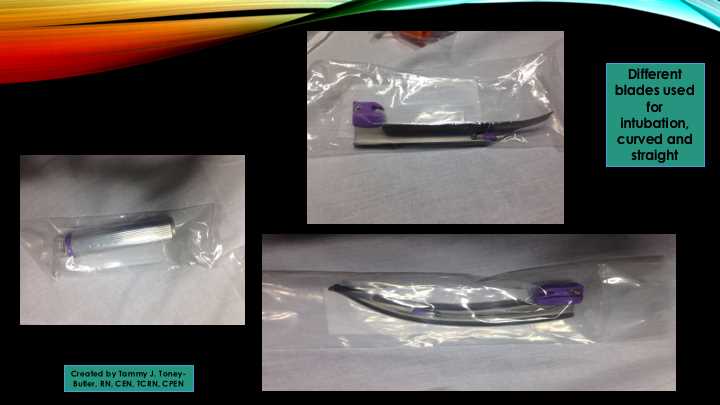
Blades and equipment needed for endotracheal intubation, curved and straight blades, disposable examples
Contributed by Tammy J. Toney-Butler, RN, CEN, TCRN, CPEN