Pericardial Calcification
- Article Author:
- Nauman Khalid
- Article Author:
- Sarah Ahmad
- Article Editor:
- Evan Shlofmitz
- Updated:
- 8/24/2020 10:47:15 PM
- For CME on this topic:
- Pericardial Calcification CME
- PubMed Link:
- Pericardial Calcification
Introduction
The normal pericardium is 1 to 2 mm thick and is comprised of an outer fibrous layer and an inner serous layer (which further subdivides into a visceral layer, or epicardium, and a parietal layer). A potential space that contains approximately 15 to 35 ml of lubrication fluid separates the visceral and parietal layers. The pericardium is a rigid, avascular, fibrous sac and its primary function is minor anchoring, lubrication, preventing distention of cardiac chambers and optimizing diastolic filling.[1] Normally the pericardium lacks any calcium deposits and calcification may be a sign of underlying inflammation or a more sinister etiology. Pericardial calcification alone is generally asymptomatic; however, signs and symptoms can develop due to underlying disease processes such as constrictive pericarditis (CP). However, a significant point to keep in mind is that pericardial calcification may not be present in up to 20% of cases of CP and it may be present in the absence of constrictive physiology. Interestingly, there have been recent reports of the development of CP after cardiac transplantation, an unusual presentation, as the transplanted heart is believed to be free of any pericardial tissue.[2][3]
Etiology
Constrictive pericarditis occurs when a normal thin compliant pericardium gets replaced by a thick, calcified, non-compliant pericardium that interferes with ventricular filling. Varying degrees of pericardial thickening and calcification is present in about 80% of the cases of CP. The etiologic basis of CP in the United States (US) has evolved over the past few decades. Historically tuberculosis had been a major cause of CP in the US and accounted for almost half of the cases, but now it is encountered less frequently. Recent studies have shown major cardiac surgery and 'idiopathic' cases as being the most common etiologies followed by radiation heart disease.[4] Other important etiologies include viral pericarditis, trauma, malignancy, rheumatologic and connective tissue diseases.[5] However, tuberculosis continues to be the most common cause of CP in the developing world.
Epidemiology
For idiopathic and viral constrictive pericarditis an incidence of 0.76 cases per 1000 person-years has been reported in a series of 500 consecutive patients; this contrasts with the connective tissue disease, neoplasms, tuberculosis, and purulent CP in which the incidence was noted to be 4.40, 6.33, 31.65, and 52.74 cases per 1000 person-years, respectively.[6]
History and Physical
Pericardial calcification is generally asymptomatic; symptoms usually develop with cardiac hemodynamic compromise such as in the setting of constrictive pericarditis. Symptoms of right heart failure or low output states such as dyspnea on exertion, orthopnea, bendopnea, and fatigue may develop. Physical examination findings include hepatomegaly, ascites, peripheral edema (usually bilateral), hepatojugular reflux, Kussmaul’s sign, jugular venous distention (with prominent x and y descents), and pulsus paradoxus.
Evaluation
Chest radiography may identify pericardial calcification; however, it suffers from low sensitivity. The laboratory workup may show an elevation of liver enzymes (especially alkaline phosphatase) and creatinine. Computerized tomography (CT) provides excellent anatomic details of the heart and pericardium. The pericardial thickness on the CT scan should be less than 2 mm.[7] Although the pericardial thickness of more than 4 mm on CT scan is predictive of constrictive pericarditis up to 20% of patients with normal pericardial thickness can still develop CP.[8] Cardiac magnetic resonance imaging can be utilized not only to delineate cardiac anatomy and pericardial thickness, it can provide information on ventricular septal motion demonstrating the so-called 'septal bounce' and 'septal shudder' which is a classic finding for CP also noted on transthoracic echocardiogram (TTE).[4]
In a patient with symptoms of right-sided heart failure and suspected constrictive pericarditis, Two-dimensional (2-D) echocardiography and tissue Doppler Imaging are probably the most important tools for diagnosing CP. Two-dimensional echocardiogram demonstrates septal motion abnormalities, enhanced interventricular interdependence, and plethoric inferior vena cava. Tissue Doppler of the mitral annulus reveals increased medial early diastolic velocities (e').[4] In healthy individuals, mitral lateral e' velocity is usually greater than the medial e' velocity, but in CP due to the tethering effect of the lateral annulus to the surrounding calcified pericardium, this velocity becomes lower, a phenomenon called 'annulus reversus' and a hallmark feature of CP.[4] In some cases where the diagnosis is unclear, an invasive hemodynamic assessment with cardiac catheterization is possible. Typically, an equalization of end-diastolic pressures in all four cardiac chambers is noted. Furthermore, with the 'match-up' study, by placing high fidelity catheters in the right and left ventricle and recording pressures simultaneously, discordant respirophasic changes in the ventricular filling patterns are noted (a catheterization equivalent of ventricular interdependence, in which one ventricle fills at the expense of other depending on the respiratory cycle).[9]
Treatment / Management
Pericardial calcification in the absence of symptoms does not require any treatment. Subacute cases of constrictive pericarditis, in which there is underlying inflammation, may respond to anti-inflammatory therapy (such as colchicine, corticosteroids, and non-steroidal anti-inflammatory drugs).[3][10] Surgical pericardiectomy is the gold standard for CP and is potentially curative.
Differential Diagnosis
Constrictive pericarditis must be ruled out when pericardial calcification is present on imaging modalities. Unusual forms of CP such as transient constriction (which responds to anti-inflammatory therapy) and effusive constrictive pericarditis (developing in patients with cardiac tamponade and develop immediately after pericardiocentesis), or occult constrictive pericarditis (which manifests after a fluid challenge) must be ruled out as well [4]. Other differentials of calcification of pericardium include pericardial scarring as a result of recurrent pericarditis. Additionally, mimickers of CP include restrictive cardiomyopathy (a myocardial disease), and severe tricuspid regurgitation must be ruled out.
Prognosis
Long-term survival after pericardiectomy for constrictive pericarditis depends on the underlying etiology. In a cohort of 163 patients, idiopathic CP had the best 7-year survival (88%) followed by postsurgical (66%) and post-radiation CP (27%).[11]
Complications
Pericardial calcification in the presence of symptoms should be evaluated further as constrictive pericarditis if present could be potentially curative. Without surgical pericardiectomy, constrictive pericarditis portends a very poor prognosis due to complications associated with heart failure and the low output state. Renal failure, organomegaly, shock, and death are other potential complications.
Deterrence and Patient Education
Early recognition and diagnosis of pericardial calcification and ruling out constrictive pericarditis is imperative to ensure timely management optimal clinical outcomes.
Enhancing Healthcare Team Outcomes
Pericardial calcification in the absence of symptoms is a benign condition. However, constrictive pericarditis must be ruled out in such patients. Diagnosis of CP can be challenging and requires a multimodality imaging with an interprofessional approach involving invasive and non-invasive cardiologists, radiologists and cardiothoracic surgeons. If missed, CP carries a grave prognosis, and most patients die with medical management alone. Pericardiectomy is potentially curative and carries acceptable mid- and long-term outcomes depending on the etiology.
(Click Image to Enlarge)
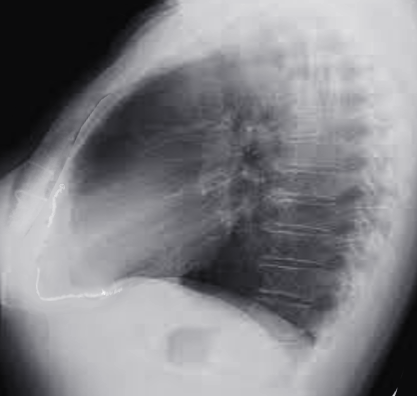
S/P CABG pericardial calcification
Image courtesy S Bhimji MD