Pityriasis Alba
- Article Author:
- Donald Givler
- Article Author:
- Hajira Basit
- Article Editor:
- Amy Givler
- Updated:
- 8/14/2020 11:07:06 PM
- For CME on this topic:
- Pityriasis Alba CME
- PubMed Link:
- Pityriasis Alba
Introduction
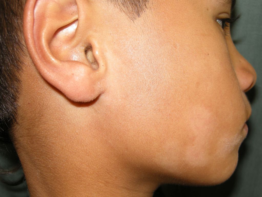
Pityriasis alba is a common, benign skin disorder occurring predominantly in children and adolescents. The name refers to its appearance: pityriasis refers to its fine scales and alba to its pale color (hypopigmentation). Most patients have a history of atopy, and pityriasis alba may be a minor manifestation of atopic dermatitis. It is characterized by ill-defined macules and patches (or thin plaques), round or oval, often with mild scaling, and sometimes with mild pruritus. The lesions initially may be mildly erythematous, and over time they become hypopigmented. They are most commonly located on the face (especially the cheeks), arms, and upper trunk, and they are more noticeable in people with darker skin types. Sun exposure accentuates the lesions. Patients and parents often are concerned about the cosmetic appearance of the lesions. Pityriasis alba resolves spontaneously, with a gradual return of normal skin pigmentation. Time to complete resolution varies from several months to a few years, although most cases resolve within one year. Reassurance, low-potency topical corticosteroids, and mild emollients are the mainstays of treatment.[1][2]
Etiology
No specific cause of pityriasis alba has been identified. It is not contagious, and no infectious etiology has been reported. It is most common in individuals with a history of atopy, although it may occur in nonatopic individuals as well. In many cases, it is considered to be a minor manifestation of atopic dermatitis. It is thought to represent nonspecific dermatitis with residual post-inflammatory hypopigmentation. Histopathology shows decreased melanin production in the affected areas. Associated findings in some studies include atrophic sebaceous glands, iron-deficiency anemia, and low levels of serum copper. The significance of these findings and their relationship to pityriasis alba is uncertain.[3]
Epidemiology
Pityriasis alba is most common in children aged three to 16 years, with 90% of cases occurring in children younger than 12 years. An estimated 5% of children in the United States may be affected. Studies have demonstrated a higher prevalence in Egypt (18%) and Mali (20%). Pityriasis alba is more common in patients with a history of atopy, and a slight male predominance has been noted. There is no clear racial predominance, although the lesions may be more noticeable in those with darker skin types. Pityriasis alba is not seasonal, although scaling may be worse in the winter (as a result of dry air in homes), and lesions may be more obvious in the spring and summer (as a result of sun exposure and darkening of the surrounding skin). Normal skin pigmentation returns spontaneously, usually within one year. [2]
Pathophysiology
The microscopic features of pityriasis alba are those of a mild, chronic, nonspecific dermatitis with decreased melanin production. Several nonspecific histopathologic features have been described. These include hyperkeratosis, parakeratosis, acanthosis, spongiosis, and perivascular infiltrates. Although there are no specific diagnostic criteria, certain features in a biopsy specimen taken from a characteristic skin lesion are suggestive of the diagnosis. These include irregular or markedly reduced melanin in the basal layer, no significant decrease in melanocyte count, and reduced number of active melanocytes with a decreased number and size of melanosomes.[4][5]
History and Physical
Although the initial lesions of pityriasis alba are mildly erythematous, the erythematous stage may go unnoticed. The most common presentation is asymptomatic (or mildly pruritic), hypopigmented lesions, often on the face. The patient or family history may include atopic dermatitis, allergic rhinitis, or asthma. The lesions may be an incidental finding on physical examination, although concern about the cosmetic appearance of the lesions on the part of the patient or the parent often leads to a consultation with a healthcare provider. The hypopigmentation often becomes more apparent with sun exposure (and darkening of the surrounding skin) during the spring and summer.
Physical examination reveals multiple round or oval-shaped hypopigmented macules or patches (or thin papules and plaques) with indistinct margins. There may be mild erythema and/or scaling. The lesions most commonly number from four to 20, measure 0.5 cm - 5 cm in size, and are distributed predominantly on the face, neck, upper arms, and upper trunk. Signs of atopic dermatitis may be present, including an eczematous rash in the popliteal or antecubital fossa, nipple eczema, cheilitis, and infra-auricular fissuring.[1][6]
It is important to carefully examine the patient because the scaly lesions may often represent psoriasis.
Evaluation
Based on the clinical appearance and distribution of the skin lesions in a child or adolescent, the diagnosis of pityriasis alba is, in most cases, straightforward.
The differential diagnosis of pityriasis alba includes postinflammatory hypopigmentation from any cause, fungal infections (tinea versicolor and tinea corporis), vitiligo, nevus depigmentosus (a stable congenital leukoderma), psoriasis, seborrhea, the ash-leaf macules of tuberous sclerosis, mycosis fungoides (cutaneous T-cell lymphoma), and hypopigmentation secondary to topical medications such as retinoic acid, benzoyl peroxide, and corticosteroids. In the proper geographic and clinical setting, the diagnosis of leprosy also should be considered.
If the diagnosis is uncertain, several diagnostic procedures may be useful. On examination with a Wood's lamp, the lesions of pityriasis alba may be accentuated but are nonfluorescent. This finding is in contrast to vitiligo, which will fluoresce more brightly and have edges with sharper demarcation. Potassium hydroxide (KOH) preparation of a skin scraping will be negative for fungal elements. This result is in contrast to tinea versicolor or tinea corporis, which will be positive for fungal elements. Skin biopsy is usually not necessary, but when performed, it can distinguish pityriasis alba from mycosis fungoides.[2][7]
Treatment / Management
Patients and their parents can be reassured regarding the benign and self-limited nature of pityriasis alba. However, they also should be made aware that its slow resolution may take several months to a few years, although most cases resolve within one year. The affected areas should be protected from sun exposure, as darkening of the surrounding skin may worsen the cosmetic appearance. Low-potency topical steroids, such as 1% hydrocortisone cream or ointment, may reduce erythema and pruritis and accelerate repigmentation. Mild emollients, such as petroleum jelly and creams, may reduce scaling. Sunscreen may help prevent the lesions from sunburning and decrease the darkening of the surrounding skin. Treatment with topical calcineurin inhibitors, such as 0.1% tacrolimus ointment and 1% pimecrolimus cream, have also been reported to be effective; however, because of their high cost, they are seldom indicated. Calcitriol, a topical vitamin D analog, showed comparable efficacy compared with tacrolimus. Other treatment options, usually reserved for extensive cases, include psoralen plus ultraviolet-A (PUVA) photochemotherapy and targeted phototherapy with a 308-nm excimer laser.[1]
Differential Diagnosis
- Atopic dermatitis
- Contact dermatitis
- Delusional tinea
- Discoid eczema
- Leprosy
- Nevus anemicus
- Nummular eczema
- Pityriasis rosea
- Seborrhea
- Tinea corporis
Prognosis
Pityriasis alba is a self-limited infection and has a good prognosis. Complete repigmentation is the norm in most people. The key issue is the cosmetic deficit for which patients often seek medical help. Also, there is a risk of sunburn in areas of hypopigmentation. The skin disorder may last a few days or a few years. In some cases, the treatment may shorten the duration of the disorder.
Pearls and Other Issues
Two uncommon variants of pityriasis alba have been described. The lesions of pigmenting pityriasis have a central zone of bluish hyperpigmentation surrounded by a hypopigmented halo. These are often associated with dermatophyte infection and are found primarily in darker skin types from South Africa and the Middle East. Extensive pityriasis alba is characterized by widespread, symmetrical, and more persistent involvement of the skin, lesions distributed predominantly on the trunk rather than on the face, a higher female-to-male ratio, and the histologic absence of spongiosis.[1][8][9]
Enhancing Healthcare Team Outcomes
Pityriasis is a common skin disorder which is best managed by an interprofessional team, because of the huge differential diagnosis.
Pityriasis alba is a common skin disorder managed by the primary care provider, nurse practitioner, dermatologist, and specially trained nurses.
The key is to educate the patient on the benign nature of the disorder. No treatment is required, and the condition spontaneously regresses in 12-24 months. For those who seek treatment for cosmesis, referral to a dermatologist is recommended. Many treatments are available, but there is no evidence to suggest that one is better than the other. Moreover, some treatments are also more likely to cause more harm than good. [1] Some patients may benefit from a moisturizer. Dermatology nurses are often involved in patient education, arrange for follow up, and facilitate communication within the team. Pharmacists can assist in the selection of emollient creams and sunscreens, also participating in patient education.
Patients should be told to avoid the sun and wear a wide-brimmed hat and long-sleeved garments. Sometimes the UV light can worsen the scaling.
The outcomes in most patients are excellent, but the recovery may take months or years.
(Click Image to Enlarge)
Pityriasis Alba
Contributed by DermNetNZ