Porokeratosis
- Article Author:
- Grant Williams
- Article Editor:
- Eric Fillman
- Updated:
- 8/11/2020 11:36:18 AM
- For CME on this topic:
- Porokeratosis CME
- PubMed Link:
- Porokeratosis
Introduction
Porokeratosis is an uncommon dermatologic disorder. It is a disorder of keratinization that presents with keratotic papules or annular plaques with an elevated border.[1] It has a distinct histologic hallmark of cornoid lamella, which is a column of tightly fitted parakeratotic cells in the upper epidermis.[2][3] There are multiple different clinical variants of porokeratosis, including disseminated superficial actinic porokeratosis, classical porokeratosis of Mibelli, porokeratosis palmaris plantaris et disseminatum, and linear porokeratosis.[2] Additionally, more rare variants include genitogluteal porokeratosis, facial porokeratosis, giant porokeratosis, porokeratosis ptychotropica, hypertrophic verrucous porokeratosis, eruptive pruritic papular porokeratosis, follicular porokeratosis, and reticulate porokeratosis.[1][4] Variants can occur together,[5] but rarely do.[1] Porokeratosis is a precancerous lesion that can undergo malignant transformation.[6] Evaluation of porokeratosis is best with biopsy of the elevated border. While multiple therapies exist for porokeratosis, including topical, systemic, and surgical, there are no standard guidelines for treatment.[7]
Etiology
The etiology of porokeratosis is unclear at this time.[3]
Epidemiology
Porokeratosis is an uncommon diagnosis. It usually occurs on sun-exposed skin, most commonly in the fifth decade of life, but can occur at any age and similar frequencies in males and females.[2] The increased occurrence of disseminated superficial actinic porokeratosis on sun-exposed skin likely indicates that ultraviolet light is a risk factor.[8]
The eruptive form of porokeratosis correlates with immune suppression, transplantation patients, inflammatory states, and malignancy.[4]
Porokeratosis will progress to nonmelanoma skin cancer in 6.9% to 30% of cases, most frequently squamous cell carcinoma, and less frequently basal cell carcinoma.[6]
While porokeratosis is usually an acquired disease, there is often a familial predisposition that could signify a hereditary component.[5]
Pathophysiology
Porokeratosis is an entity that results from the disordered progression of epidermal cells.[9] The development of this entity can be related to sun exposure. In areas where there has been no or limited sun exposure, repeated minor frictional trauma due to tight clothing may cause the entity, as is the case in genitourinary porokeratosis.[1]
There is a reported association with overexpression of p53, and occasionally there can be an expansion of a clone of abnormal epidermal keratinocytes.[6][5] Porokeratosis has the possibility of malignant transformation into squamous cell carcinoma or basal cell carcinoma.[5]
Histopathology
The distinctive histopathologic feature of porokeratosis is a cornoid lamella, which is a column of tightly fitted parakeratotic cells.[2][3] The column of parakeratosis will occur over the epidermis with an absent granular layer and dyskeratotic cells in the upper spinous zone.[1] This feature is usually present at the elevated border of the lesion.[1] While once thought to be pathognomonic of porokeratosis, the feature has presented in other conditions.[9] Cornoid lamellation reflects a disordered progression of epidermal cells.[9] In follicular porokeratosis, the cornoid lamella can involve the follicular infundibulum.[10]
History and Physical
Porokeratosis presents as keratotic papules or annular plaques that expand centrifugally with an elevated keratotic border.[1] Centrally, the lesion can appear slightly atrophic.[1] The lesions can present with pruritus and may be present for several years before diagnosis.[1] Disseminated nodules present as pink to brown papules and macules with raised borders.[4] Porokeratosis most commonly occurs in the limbs and trunk but can occur in the trunk, face, and genitourinary region, and scrotum.[10] However, porokeratosis as an entity has a broad spectrum of presentations, including large destructive lesions and involvement of burn scars.[6]
Evaluation
Evaluation of porokeratosis is best with a biopsy of the elevated border.[1] Biopsy in this area will show the cornoid lamella and will be essentially diagnostic. Dermoscopy can also be useful for diagnosis. Dermoscopy of lesions shows central brown pigmentation with blue-gray dots surrounded by a single hypopigmented band with a white track at the periphery.[1]
Despite the relative rarity of porokeratosis, especially some subtypes like genitourinary porokeratosis, one should keep the diagnosis in mind due to the risk of malignant degeneration.[6]
Treatment / Management
Multiple therapies are described for porokeratosis, including topical, systemic, and surgical. However, there have been no randomized controlled trials, so there are no international guidelines on treatment standards.[7]
- Classical porokeratosis of Mibelli is most successfully treated with imiquimod cream.[7]
- Linear porokeratosis responds well to topical or systemic retinoids.[7]
- Disseminated porokeratosis can be treated successfully with topical vitamin D acid derivatives.[7]
- Surgical interventions or cryotherapy are options for treatment in areas where topical agents are challenging to use or contraindicated.[7] Laser therapy may be another treatment option.[4]
- Topical steroids, retinoids, and topical diclofenac may provide symptomatic relief even if no lasting benefit occurs.[1]
Differential Diagnosis
Clinically porokeratosis can resemble psoriasis, lichen simple chronicus, hypertrophic lichen planus, tuberculosis of the skin, and Bowen disease, candidiasis, irritants, and allergic contact dermatitis.[1]
Prognosis
Porokeratosis is a premalignant condition[1]; however, all types of porokeratosis can undergo malignant transformation to non-melanoma skin cancer at a rate of 6.9 to 30%.[2][6] Most commonly, the lesion transforms into squamous cell carcinoma. Less frequently, the lesion transforms into basal cell carcinoma. If the lesion has not undergone a malignant transformation, excision is curative.
Linear porokeratosis and giant porokeratosis are the variants that are most susceptible to malignant transformation. Malignant transformation is most rare in disseminated superficial actinic porokeratosis.[11]
Deterrence and Patient Education
Patients should use sun protection, avoid excessive sunlight exposure, and go for periodic examination by a dermatologist to monitor for recurrence or malignant transformation.[2]
Pearls and Other Issues
- The hallmark of porokeratosis histologically is a cornoid lamella, which is a column of tightly fitted parakeratotic cells.[3]
- Porokeratosis presents as keratotic papules or annular plaques that expand centrifugally with an elevated keratotic border.[1] Dermoscopy of lesions shows central brown pigmentation with blue-gray dots surrounded by a single hypopigmented band with a white track at the periphery.[1]
- There are many variants of porokeratosis.[2]
- Porokeratosis can undergo malignant transformation to squamous cell carcinoma and basal cell carcinoma.[6]
- Evaluation of porokeratosis is best done with a biopsy of the elevated border.[1]
- Multiple therapies are described for porokeratosis, including topical, systemic, and surgical. However, there have been no randomized controlled trials, so there are no international guidelines on treatment standards.[7]
- Patients treated for porokeratosis should be advised to use sun protection, avoid excessive sunlight exposure, and go for periodic examination by a dermatologist to monitor for recurrence or malignant transformation.[2]
Enhancing Healthcare Team Outcomes
Dermatologists and other clinicians should work closely with pathologists to ensure lesions receive an evaluation for the presence of malignant transformation.[2] [Level 5]
Dermatologists and dermatology specialty nurses should periodically followup with patients to monitor for recurrence.[2] An interprofessional team approach to follow up will provide the best patient outcomes. [Level 5]
(Click Image to Enlarge)
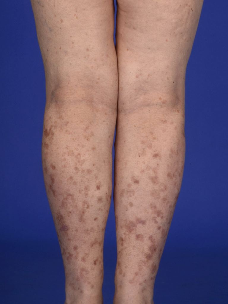
Disseminated Superficial Actinic Porokeratosis (DSAP)
Contributed by DermNetNZ
(Click Image to Enlarge)

Porokeratosis
Contributed by DermNetNZ
(Click Image to Enlarge)
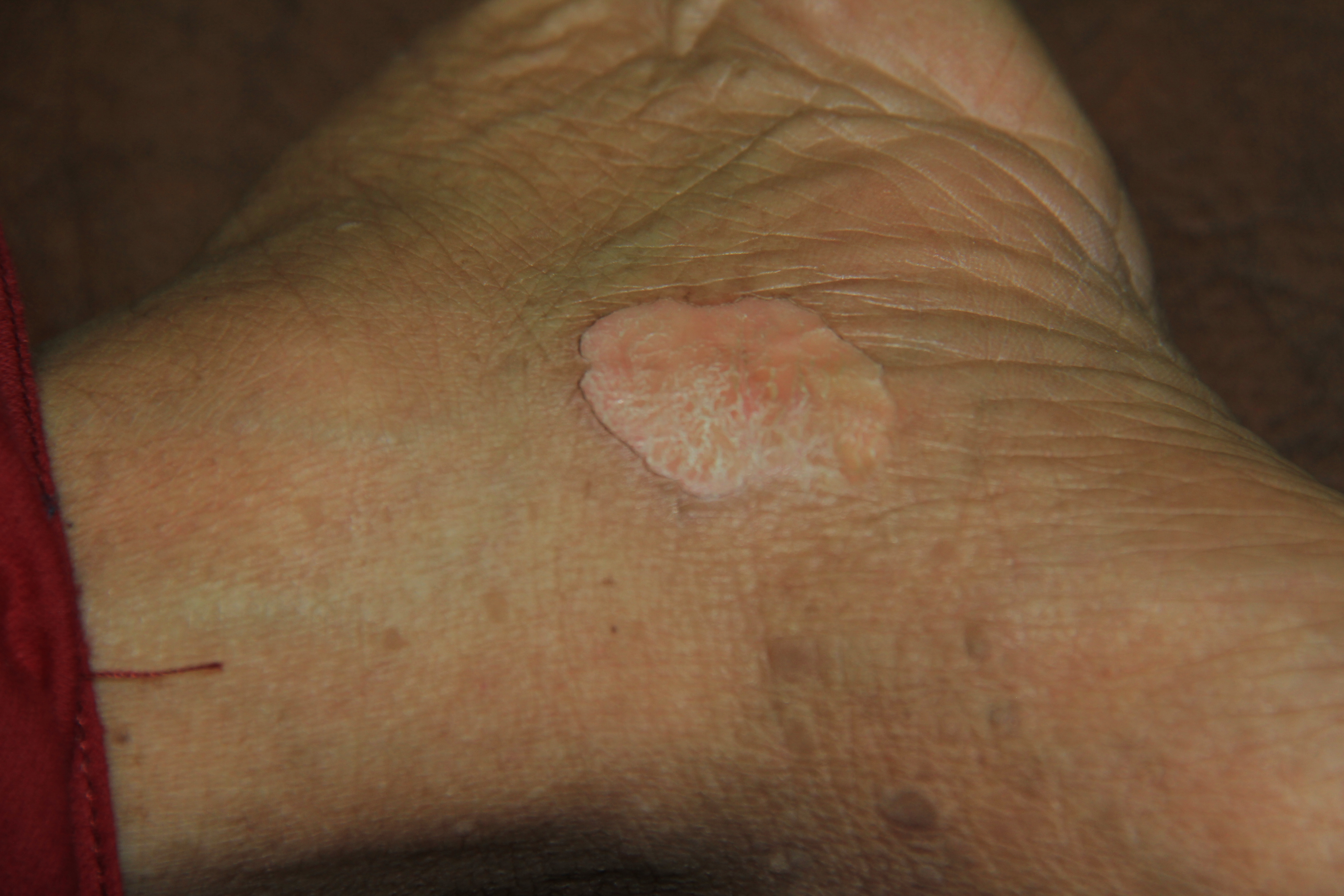
Porokeratosis of Mibelli
Contributed by Dr. Shyam Verma, MBBS, DVD, FRCP, FAAD, Vadodara, India