Ptosis
- Article Author:
- Babar Shahzad
- Article Editor:
- Marco Siccardi
- Updated:
- 8/10/2020 5:44:14 PM
- For CME on this topic:
- Ptosis CME
- PubMed Link:
- Ptosis
Introduction
Ptosis is abnormally low positioned upper eyelid, also called blepharoptosis, which can decrease or even occlude the vision completely. It may be congenital or acquired in origin. Proper management requires recognizing the exact etiology and treat it accordingly, whether surgically or medically, to improve patient outcome.[1]
Etiology
Ptosis is classified into congenital or acquired based on the age of presentation; the latter is usually further categorized into five types based on etiology[2]:
- Neurogenic: It results from defective innervation of the levator muscle of upper eyelid. For example, third nerve palsy, Horner syndrome, Marcus Gunn jaw-winking syndrome, multiple sclerosis, etc.
- Myogenic: Levator muscle myopathy or defect at its neuromuscular junction causes the myogenic ptosis which includes myasthenia gravis, ocular myopathy, simple congenital, blepharophimosis syndrome, etc.
- Mechanical: Levator function bis impaired due to the mass effect of some abnormal external structure such as neoplasm, chalazion, contact lens in the upper fornix, scarring, etc.
- Aponeurotic: Also known as Involutional ptosis, it results from a defective levator aponeurosis due to aging, trauma, or postoperative complication.
- Traumatic: Any kind of direct or indirect trauma to the eyelid leading to levator transection, cicatrization, eyelid laceration or orbital rooftop fracture with ischemia can cause ptosis.
Epidemiology
Among all cases of ptosis, congenital ptosis is the most common type which seems to be more prevalent in males. Simple congenital ptosis is the most prevalent form of congenital ptosis.[3] Among acquired cases, aponeurotic ptosis is the most common type which usually presents in late adulthood.[4] Enough data is not available yet, about the incidence of ptosis.[5] However, the prevalence of ptosis does not seem to be affected by other epidemiological factors such as race, etc.
Pathophysiology
Levator palpebrae superioris (LPS) and Muller's muscle are two muscles of upper eyelid responsible for its elevation. LPS is the main elevator which is supplied by the oculomotor nerve. The levator palpebrae superioris muscle origin is the lesser wing of the sphenoid bone, it travels anteriorly above the superior rectus muscle, and attaches in multiple insertions: anteriorly into the upper eyelid skin, inferiorly on the anterior surface of the upper tarsal plate and to the superior conjunctival fornix. Muller's muscle is a smooth muscle also attached to the superior tarsal plate with sympathetic innervation, which is defective in ptosis of Horner syndrome.[1] Loss of innervation of LPS and Muller's muscle causes neurogenic ptosis.
Levator muscle dystrophy causes simple congenital ptosis. On the other hand, involutional changes in the eyelid are the most common pathogenesis in adult ptosis. Deceasing tone and thinning of the levator muscle, due to aging, results in abnormal position of the eyelid. Disinsertion of the levator aponeurosis, or its dehiscence, after any trauma or surgery, can also lead to ptosis.[5]
History and Physical
Presentation:
Main presenting complain of patients with ptosis is the visual disturbance ranging from mild to severe, which can be unilateral or bilateral along with cosmetic disfigurement. Ptosis may accompany with some other problems depending upon the etiology.
History:
Age of onset and duration of symptoms is important to differentiate congenital from acquired cases. There also may be other symptoms of associated etiologies, such as diurnal variability, diplopia, eyeball deviation, body fatigability, etc. The patient should be asked about any previous history of trauma, surgery, or medical treatment.[6]
Clinical Evaluation
In the case of ptosis, the upper lid margin covers more than 2 mm of the cornea, making palpebral fissure narrower than normal. The examiner should observe both eyes and the general appearance of the patient. Backward tilted head, wrinkled skin of the forehead, and elevated eyebrows may present as compensatory changes.[2] Any scar, swelling, or abnormal structure near eyelids should not be missed. It may present with eyeball deviation, shrinking or bulging. Following clinical signs should also be examined:
- Absent upper lid crease signifies the congenital ptosis.
- Pupillary function, i.e., anisocoria in Horner syndrome (miosis) and CN III palsy (mydriasis).[7]
- Ocular motility to assess CN III paresis.
- Jaw-winking sign to rule out Marcus Gunn jaw-winking syndrome.
- Bells phenomena and tear film are checked preoperatively to assess fitness for ptosis surgery.[2][8]
- Phenylephrine test is used to assess the Muller's muscle before conjunctival resection. Topical phenylephrine (alpha-adrenergic sympathomimetic) is administered in the superior fornix, and eyelid elevation in response shows ideal candidacy for this surgery.[1]
Myasthenia gravis can manifest as generalized or solely ocular disease with pupil sparing ptosis. It can be assessed by edrophonium test which contains edrophonium, a short-acting acetylcholinesterase inhibitor. A positive test is the eyelid elevation in 2 to 5 minutes after administration of edrophonium.[9] It has relatively low sensitivity, and the ice test has superseded it; in which an ice pack is placed over the ptotic lid for 2 to 5 minutes and then improvement is noticed.[10]
Evaluation
Proper diagnosis and management of blepharoptosis require assessment by the following measurements:
- Levator muscle function: Assessment of the functional status of the levator muscle is by placing the thumb firmly against the patient’s eyebrow, with eyes in downward gaze. Then patient looks upward, and the amount of excursion is measured with a scale which can be graded as normal (15 mm), excellent (over 12 mm), good (9 to 11 mm), fair (5 to 9 mm) or poor (less than 4 mm).[11] This test is very important in determining the surgical procedure of choice for ptosis correction.
- Margin-reflex distance: Patient fixates on torchlight held by the examiner and the distance between its corneal reflection and the upper lid margin is measured. A value of 4 to 5 mm is normal MRD.
- Palpebral fissure height: Normally, the upper lid margin covers 2mm and lower lid margin covers 1mm of the cornea. Distance between the two is measured in the pupillary plane; 7 to 10 mm in males and 8 to 12 mm in females are normal. Comparing it with the contralateral side and calculating the difference is used to quantify the unilateral ptosis as mild (1 to 2 mm), moderate(3 to 4 mm) or severe(4 mm or more).[2]
- Margin crease distance: MCD is the distance between the lid margin and skinfold of upper lid measured in downward gaze. Normal values are 7 to 8 mm in males, and 8 to 10 mm in females is considered normal. It is higher than normal in aponeurotic ptosis, whereas absent or vague in congenital ptosis.[1]
If myasthenia gravis is suspected, blood serum is tested for acetylcholine receptor antibodies which are responsible for this autoimmune disorder. However, these are positive only in 50% of patients with solely ocular myasthenia.[9] Antistriated muscle antibodies and muscle-specific tyrosine kinase levels are also checked when myasthenia is highly suspected.
Thyroid studies are not needed usually, but myasthenia often correlates with thyroid disorders and rarely ptosis may present in hypothyroidism.[12] Thus, whenever thyroid involvement is suspected, thyroid functions must be assessed.
Imaging studies such as X-ray or brain and orbit CT/MRI scans are needed by ophthalmologists and neurologists when any pathology is suspected in these regions such as a tumor in orbit or skull, nerve defects, multiple sclerosis, trauma, etc. Thorax radiography is used to assess the thymus in case of myasthenia.
Treatment / Management
Treatment of ptosis depends upon the underlying etiology, the degree of ptosis, and the function of the levator muscle. In mechanical ptosis, removal of the abnormal structure, i.e., a chalazion, is all that is needed. However, surgical correction is the mainstay of treatment as well as some nonsurgical options available for specific conditions.
- Surgical treatment: Surgery is necessary for congenital, ptosis, and all other types when nonsurgical treatment is not beneficial: the underlying cause and preoperative evaluation of ptosis help in determining the procedure of choice.
- Levator resection: Levator muscle gets shortened by resecting the muscle if it is not paralyzed completely with mild (2 mm) to moderate (3 to 4 mm) ptosis.[13] There are different approaches for this purpose:
- Everbursch: Approach through the skin.
- Blaskovics: Approach through palpebral conjunctiva.
- Fasanella-Servat: A portion of the tarsal plate, palpebral conjunctiva and Muller's muscle get excised along with levator resection. It is usually a proposed option in minimal ptosis.[2]
- Motais procedure: Action of superior rectus is utilized to elevate the lid if levator muscle is paralyzed.
- Hess's procedure: Frontalis muscle is used to raise the lid if both, the superior rectus and the levator muscle, are paralyzed.
- Frontalis brow suspension: Eyelid is tethered with the frontalis muscle by either fascia tunica obtained from fascia lata or some other suitable synthetic material such as mersiline mesh. It is indicated in severe (over 4 mm) ptosis with poor levator function, especially in the case of congenital ptosis.[14]
- Aponeurotic strengthening: It involves the advancement of aponeurosis, mostly indicated in aponeurotic disinsertion or involutional ptosis.[13]
- Levator resection: Levator muscle gets shortened by resecting the muscle if it is not paralyzed completely with mild (2 mm) to moderate (3 to 4 mm) ptosis.[13] There are different approaches for this purpose:
- Nonsurgical treatment: It is indicated in myogenic and some cases of neurogenic ptosis mostly. For example, taping upper lid open and spectacle props (eyelid crutches attached to glasses).
Differential Diagnosis
Pseudoptosis is the false perception of the ptosis, which may result from the following[1][2][6]:
- Contralateral retracted lid
- Downward deviated ipsilateral eyeball followed by the upper lid
- Overhanging skin over the upper lid
- Brow ptosis
- Upper lid swelling, i.e., preseptal cellulitis, orbital cellulitis, chalazion, etc.
- Volume deficit as seen in microphthalmos, phthisis bulbi, enophthalmos, etc.
Prognosis
Prognosis depends upon the type and treatment of ptosis. The appropriate surgical procedure applied according to the preoperative evaluation shows very good prognosis. For example, ptosis with poor levator function corrected with the levator resection instead of fascia lata sling procedure will show poor results. Proper preoperative assessment and postoperative care with close monitoring fairly improve the outcome of surgical repair in any form of ptosis.
Complications
Complications that can occur after ptosis repair can subdivide into:
- Early complications: Postoperative asymmetry in eyelid height and shape is the most common complication.[15] Under correction or overcorrection commonly appears postoperatively, which may resolve with time. Surgical site infection and bleeding from the wound are acute but uncommon complications followed by exposure keratopathy, which requires proper monitoring. Postoperative mild lagophthalmos or incomplete closure of palpebral fissure is common, which usually resolves within 2 to 3 weeks.[2]
- Late complications: Persistent lagophthalmos, exposure keratopathy, and recurrent ptosis are chronic complications of ptosis repair surgery. Persistent under correction or overcorrection require revision surgery.[1]
Deterrence and Patient Education
Patient education regarding the cause of ptosis and long-term complications it can cause if untreated is necessary. All treatment options, surgical and nonsurgical, including the best one according to assessment, should be discussed. The clinician must explain likely postoperative complications to the patient that can occur earlier or later on and their management.
Enhancing Healthcare Team Outcomes
Ptosis is one of the most commonly presenting cases in ophthalmology clinics, which requires management with coordination between the ophthalmologist, optician, nurse, and the surgeon for proper management. The majority of patients with ptosis first present in primary care clinics, and it is important to refer these patients to a neurologist and/or ophthalmologist for further workup.
This teamwork is vital for the preoperative assessment and choosing the best treatment option for a particular case of ptosis. Surgical correction of ptosis should be carried out by an oculoplastic surgeon along with a plastic surgeon for the best visual as well as the cosmetic outcome. Specialty-trained nursing will prep the patient, assist during the surgery, and provide post-surgical care and counsel to the patient and/or family, reporting any concerns to the surgeon or other physicians managing the case. Knowledge and evaluation of the case are crucial for the surgeons to perform a procedure in the best interest of the patient. This type of interprofessional collaboration is vital to achieving optimal patient outcomes in ptosis cases. [Level 5]
(Click Image to Enlarge)
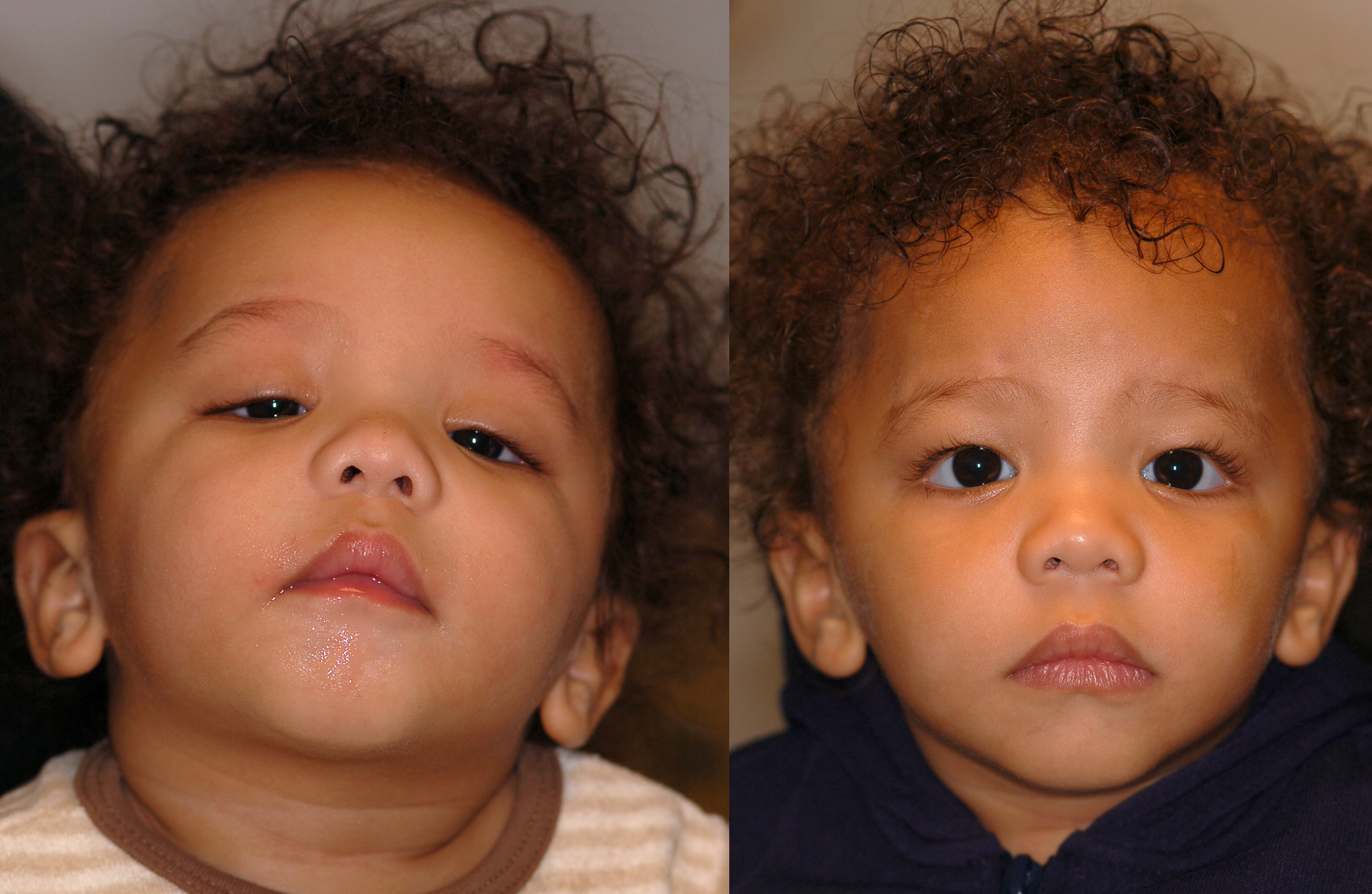
Patient with levator function less than 4 mm bilaterally with a preoperative chin-up position and severe ptosis. Bilateral frontalis slings (using polytetrafluoroethylene this child) performed to lift the upper eyelids utilizing the brows.
Contributed by Prof. BCK Patel MD, FRCS