Purtscher Retinopathy
- Article Author:
- Koushik Tripathy
- Article Editor:
- Bhupendra Patel
- Updated:
- 8/11/2020 11:37:34 AM
- For CME on this topic:
- Purtscher Retinopathy CME
- PubMed Link:
- Purtscher Retinopathy
Introduction
Purtscher retinopathy (traumatic retinal angiopathy or lymphorrhagia retinae or retinal teletraumatism) is an occlusive microvasculopathy characterized by multiple retinal white areas around the optic nerve head and fovea with paravascular clearing which may be associated with intraretinal hemorrhages. Purtscher flecken, cotton wool spots, and minimal intraretinal hemorrhage are typical features. Management depends on the cause, and the role of systemic steroid need further evaluation.
Typical Purtscher retinopathy described by Otmer Purtscher has features like severe visual decline after head trauma. In the initial report, it was noted to be associated with multiple superficial retinal white patches, retinal hemorrhages, and disc edema.
When features like Purtscher retinopathy presents without the history of frank trauma, it is called Purtscher-like retinopathy.
Etiology
Causes of Purtscher retinopathy are associated with trauma/surgery. These include[1]:
- Severe head trauma
- Chest compression
- Fracture of long bones (eg. femur) or crush injury
- Dislocation of the shoulder joint and avulsion fracture of the greater tuberosity of the humerus
- Barotrauma
- Battered baby syndrome
Causes of Purtscher-like retinopathy include[2][3][4][5][6][7][8][9][10][11][12][13][14][15][16][17][18][19][18][17][16][15][14][13][12][11][10][9][8][7][6][5][4][3][2]:
- Acute pancreatitis
- Chronic pancreatitis
- Pancreatic adenocarcinoma
- Embolism (fat, air, amniotic fluid)
- Chronic renal failure
- Severe acute retinal failure associated with squamous cell carcinoma of the cervix
- Acute renal allograft rejection
- Nephrotic syndrome
- Connective tissue disorder (systemic lupus erythematosus, scleroderma, dermatomyositis)
- Thrombotic thrombocytopenic purpura (TTP)
- Cryoglobulinemia in hepatitis C
- Hemolytic uremic syndrome (HUS), atypical hemolytic uremic syndrome
- Hemophagocytic lymphohistiocytosis
- Preeclampsia
- HELLP syndrome (hemolysis, elevated liver enzymes, low platelets)
- Eclamptic posterior reversible encephalopathy syndrome
- Placental abruption
- Valsalva maneuver
- Weight lifting
- Retrobulbar and peribulbar anesthesia
- Steroid injection around the orbit
- Hodgkin lymphoma after bone marrow transplant and antineoplastic therapy as a result of retinal thrombotic microangiopathy, relapsing Hodgkin lymphoma treated with brentuximab
- Hematopoietic stem cell transplant-associated thrombotic microangiopathy
- Multiple myeloma
- Leukemia
- After childbirth which may be associated with amniotic fluid embolism and disseminated intravascular coagulation
- Iron deficiency anemia
- Hypersensitivity to drugs or anaphylaxis after drug use
- Gemcitabine in a diabetic patient with small cell lung cancer with necrotizing vasculitis resulting in necrosis of the distal finger. This patient also had elevated antinuclear antibody
- Acute myocardial infarction
- Recent viral illness or acute febrile illness or dengue fever
- After coil embolization of intracavernous carotid artery embolism associated with Horner syndrome in the ipsilateral eye or coil embolization of middle cerebral artery aneurysm
- Primary hypereosinophilic syndrome
- Adult-onset Still disease with thrombotic microangiopathy
- Pemphigus vulgaris
- After radical prostatectomy
- Endonasal dacryocystorhinostomy
- Major vascular surgery (cerebrovascular or cardiovascular)
- “Brazilian booty" retinopathy after polymethyl methacrylate (PMMA) injection into the buttock for cosmetic enhancement
- Orthopedic surgery
Epidemiology
The annual incidence of symptomatic Purtscher's retinopathy has been estimated to be 0.24 cases per million population per year.[20] However, as many cases are asymptomatic, the actual incidence of Purtscher retinopathy may be higher.
Pathophysiology
The retinal appearance is thought to be due to occlusion of peripapillary terminal arterioles which supply the superficially peripapillary capillaries.[21]
Leukoembolization, endothelial damage, activation of complement C5 and blockade of small arterioles causing infarct of the capillary bed have been implicated in the pathogenesis of Purtscher retinopathy and Purtscher-like retinopathy. C5a component of the complement predisposes to the aggregation of leukocytes (granulocytes) which embolize. These microemboli are very small (60 to 80 microns) and cause multiple small arteriolar occlusion, instead of causing occlusion at the bifurcation of major arterioles which cause branch retinal arteriolar occlusion. In the animal model, intraarterial injection small glass balls (ballotini, 15 to 40 microns) into the external carotid artery has been shown to cause similar lesions in retina and occlusions of retinal arterioles.[22]
Occlusion of the retinal capillary bed causes Purtscher flecken, which are typical of the disease. Infarcts of the nerve fiber layer are known as cotton wool spots or soft exudates. As the area of the retina around the vessels is free of capillaries, paravascular clearing is also a feature. Another explanation of paravascular clearing is high oxygen saturation near the vessels.
Other sources of emboli include:
- Amniotic fluid embolism during childbirth or after birth
- Fat embolism after the fracture or crush injury of large bones. Acute pancreatitis may also cause fat embolism from enzymatic degradation of omental fat. Fat embolism may be present in 5% of patients with fracture of long bones, and up to 60% of patients with fat embolism have retinal manifestations. Features of fat embolism include respiratory distress, confusion/agitation, petechial rashes, and tachycardia.
- Air embolism from chest compression injury after various causes including road traffic accident
- Fibrin, platelet
Points in favor of retinal arteriolar embolism include multifocal lesions, sudden onset, normal appearing retinal arterioles which show occlusion on fluorescein angiogram, and geographic distribution of ischemic patch.
Originally, in the first reports, the proposed pathomechanism was hypothesized to be due to extravasation from lymphatic vessels secondary to a sudden rise of the intracranial tension.
Other proposed mechanisms include[23][24][23]:
- Rheological disturbances along with downstream endothelin-induced vasculopathy
- In chest compression, reduced venous return and acute expansion of retinal veins may lead to a similar retinal appearance
- Vascular (venous or arteriolar) damage from increased intravascular pressure
- Vasculitis due to free fatty acids
- Thrombotic microangiopathy categorizes a group of disorders characterized by hemolytic anemia (with prominent red blood cell fragmentation), thrombocytopenia, and thrombosis in the microvasculature. It is a feature in TTP, HUS, and various other diseases. Reduced activity of a von Willebrand factor-cleaving protease ADAMTS-13 correlates with the pathogenesis of thrombotic microangiopathy. Thrombotic microangiopathy is important in the pathogenesis of Purtshcer-like retinopathy in many cases.
Histopathology
Histopathological evaluation of Purtscher-like retinopathy in a 32-year-old black woman with pancreatitis revealed 'arteriolar occlusions in both the choroid and retina and inner ischaemic infarctions with inner retinal' edema.[25] However, in another report of Purtscher-like retinopathy in acute necrotizing pancreatitis, an 'absence of vascular occlusion and endothelial damage' was noted.[26] The vessels in and around the cotton wool spots were intact. The authors suggested that vascular embolization was not mandatory for the development of cotton wool spots in Purtscher-like retinopathy in acute pancreatitis.[26]
History and Physical
Usually, in Purtscher retinopathy, the patient presents with sudden onset painless visual decline in both eyes within 2 days after trauma. The severity of retinal involvement does not correlate with the severity of chest trauma.
Also, in the absence of trauma, Purtscher-like retinopathy may be seen in multiple disorders as described in the etiology. A detailed history is of utmost importance, and specifically, pancreatitis and systemic lupus erythematosus should be ruled out. The disease is bilateral in 60% of cases.[27] Apparently, unilateral cases may have subtle changes in the fellow eye. There is no direct ocular trauma, and clinically no arteriolar embolus is visible.
A diagnostic criterion of Purtscher retinopathy has been suggested. Three out of the following five criteria should be present.
1. Purtscher flecken
2. Retinal hemorrhages, low-to-moderate number (1 to 10)
3. Cotton-wool spots (typically restricted to posterior pole)
4. Probable or plausible explanatory etiology
5. Complementary investigation compatible with the diagnosis
Retinal zones have been used to describe the involvement in Purtscher retinopathy.[20]
- Zone A - Horizontally extends four disc diameters on either side of the fovea
- Zone B - The outer margin is the equator.
- Zone C - This is the outermost zone and extends till the ora serrata.
A broad case definition was part of a large study on Purtscher retinopathy.[20] The definition included varying combinations of the following-
(1) An associated contributing illness such as acute pancreatitis, long bone fracture, orthopedic surgery, chest compression or crush injury(2) Multiple areas of polygonal retinal whitening between the retinal arterioles and venules (Purtscher flecken) and/or superficial cotton wool spots in one or both eyes
- Typically restricted to the posterior pole
- Accompanied by minimal, if any, retinal hemorrhage
- No visible emboli in the large retinal vessels
- No direct ocular trauma
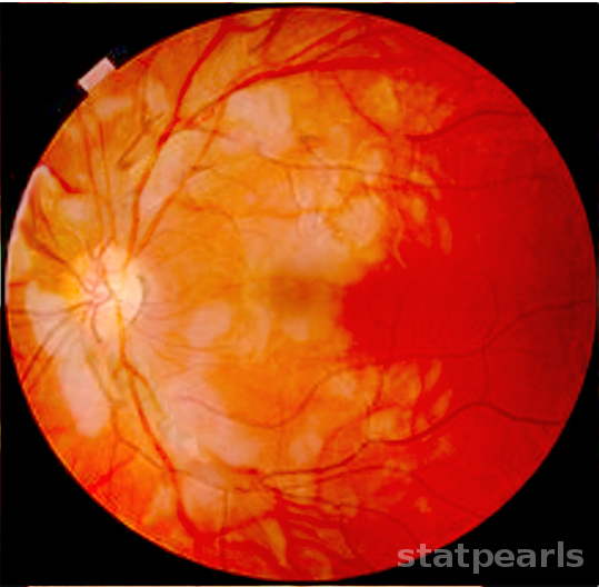
Purtscher flecken characteristically presented by multiple polygonal patches of intraretinal whitening at the posterior pole and around the optic disc. There is a clear zone of 50 microns on either side of the retinal vessels (arterioles or venules[27]). In some cases, the whiteness may reach near the venules, though a clear zone around arteriole is usually present.[28] This situation is seen in around half of the patients, though the flecken is considered pathognomonic of Purtscher retinopathy. A pseudo-cherry-red spot may present at the macula.
Cotton wool spots are whitish superficial mild elevated lesions of the inner retinal surface oriented along with the retinal nerve fiber layer. The margin of these lesions are blurred and are superficial to retinal vessels.
Other features include disc edema, macular edema, macular ischemia/infarction, and arterial occlusion. Serous macular detachment is usually a feature in pregnancy-induced hypertension. Preretinal hemorrhage may is a rare feature.[17]
Visual acuity may be 20/20 to finger counting. The patient may complain of the central or paracentral scotoma.
Evaluation
Ocular investigations include
Fundus fluorescein angiogram (FFA) - FFA features may vary. The findings include blockage of choroidal fluorescence by the whitened opaque retina, macular ischemia/infarction, capillary nonperfusion, focal areas of arteriolar occlusion, paravascular staining and leakage from the optic nerve head. Indocyanine green angiogram revealed choroidal hypocyanescence, which persisted at 5 months' follow up after trauma in a patient with Purtscher retinopathy - this may contribute to poor final vision noted in some of these cases.[29]
Optical coherence tomography (OCT) - OCT in acute phase reveals inner retinal hyperreflectivity at the Purtscher flecken and cotton wool spots. Intraretinal and subretinal fluid and retinal thickening may be associated. Paracentral acute middle maculopathy (PAMM) characterized by hyperreflectivity of the inner nuclear layer due to ischemia of intermediate and deep retinal capillary plexus may be a feature in the acute phase. In the late phase, retinal thinning, and photoreceptor loss is present.[30]
OCT angiography may show ischemia at both retinal capillary plexus which may recover, leading to visual improvement.[31]
Multifocal ERG revealed dysfunction of both inner and outer retina in a case.[32]
Visual field may reveal central or paracentral or arcuate scotoma usually with preservation of the peripheral visual field.[28]
Evident history of severe trauma is present in Purtscher retinopathy. In cases of Purtscher-like retinopathy, investigations may include
- Amylase and lipase level to rule out pancreatitis
- Imaging for injury as required
- Antinuclear antibody - Lupus retinopathy appears very similar to Purtscher-like retinopathy and should be ruled out when there is no apparent history of trauma present.
- Complement component C5 is usually elevated in Purtscher-like retinopathy and play an essential role in leukoembolization.
- Abdominal imaging to rule out pancreatitis, pancreatic cancer
- Evaluation of connective tissue disorders including
- Anti-double-stranded DNA
- Antiphospholipid antibody
- Rheumatoid factor
- Markers of muscle breakdown in dermatomyositis including serum creatine phosphokinase and serum/urine myoglobin
Treatment / Management
The management of Purtscher retinopathy consists of managing the cause and supportive therapy.[33] A systematic review did not find any difference in the improvement of visual acuity when they compared treatment with steroid with observation.[27] However, there are reports of visual and anatomical improvement with the use of three daily doses of intravenous methylprednisolone pulse (1000mg) followed by oral steroids. The proposed mechanism of action includes stabilization of the microvasculature and neuronal membrane and inhibition of granulocyte aggregation.
Cases with macular edema may benefit from anti-vascular endothelial growth factor agents like bevacizumab.[34] Lupus retinopathy, a common differential for Purtscher-like retinopathy signifies systemic activity of systemic lupus erythematosus. Such cases require systemic steroid (oral with or without intravenous pulse methylprednisolone) and immunosuppression.
A case of the atypical hemolytic uremic syndrome with thrombotic microangiopathy and Purtscher-like retinopathy had successful treatment with eculizumab (a monoclonal antibody that inhibits cleavage of C5 to C5a and C5b, thereby blocking the terminal pathway of the complement system).[31]
Other therapies attempted for this retinopathy include oral nonsteroidal anti-inflammatory drugs (indomethacin),[35] and papaverine hydrochloride. Future research is needed to manage the thrombotic microangiopathy, which is the crucial factor in the pathogenesis of many cases.[33] Bortezomib, a proteasome inhibitor can deplete ADAMTS-13 antibody (which plays a vital role in thrombotic microangiopathy) in TTP.[36]
Differential Diagnosis
Differential diagnoses of Purtscher-like retinopathy include[37]:
- Hypertensive retinopathy - May have multiple cotton wool spots. However, Purtscher flecken are not present. Arteriovenous crossing changes, disc edema, flame-shaped hemorrhage, disc edema, and hypertensive choroidopathy may be a present.
- Lupus retinopathy - Systemic features of systemic lupus erythematosus may be present.
- Partial central retinal arterial occlusion
- Endogenous endophthalmitis
- Retinitis
- Giant cell arteritis
- HIV retinopathy
- Ischemic central retinal venous occlusion
- Post-febrile retinitis
- Commotio retinae
Prognosis
The visual prognosis is variable, and in severe disease, the prognosis is usually poor. Poor prognostic factors include[38][28][38]:
- Macular infarction
- A long duration of acute retinal changes
- Optic disc swelling
- Choroidal hypoperfusion
- Severe retinal capillary nonperfusion
- Prior episode of Purtscher retinopathy in the same eye
- Involvement of the outer retina
Agarwal and McKibbin reported 15 cases with Purtscher retinopathy or Purtscher-like retinopathy caused by road traffic accident with or without long bone fracture, chest compression, and acute pancreatitis.[20] Only 1 one of the 24 eyes had a final visual acuity worse than presentation. With observation alone, 23% of eyes improved by at least 4 Snellen lines and 50% eyes improved by at least 2 Snellen lines.[20]
Complications
The long term sequelae of Purtscher retinopathy or Purtscher-like retinopathy include:
- Optic atrophy/ optic disc pallor
- Retinal pigment epithelial atrophy
- Thinning of the retinal nerve fiber layer
- Foveal thinning
- Attenuation or sheathing of retinal vessels
Deterrence and Patient Education
The patients with pancreatitis should avoid alcohol. Protective measures, including seat belts, may help reduce the injury due to the road traffic accident and possibly Purtscher retinopathy.
Enhancing Healthcare Team Outcomes
Interprofessional collaboration is vital in the management of Purtscher retinopathy. The trauma may be life-threatening, and a team approach is required, which involve multiple specialties, including ophthalmology, emergency medicine, radiology, laboratory medicine, nursing, neurosurgery, orthopedics, and critical care medicine. For Purtscher-like retinopathy, the interprofessional team may need doctors from different specialization, including internal medicine, rheumatology, gynecology & obstetrics, nephrology, and others.
Purtscher retinopathy requires an interprofessional team approach, including physicians, specialists, specialty-trained nurses, and pharmacists, all collaborating across disciplines to achieve optimal patient results. [Level V]
(Click Image to Enlarge)
Purtscher Retinopathy
Image courtesy S Bhimji MD