Splinting
- Article Author:
- Alyssa Althoff
- Article Editor:
- Russell Reeves
- Updated:
- 8/14/2020 8:01:11 PM
- For CME on this topic:
- Splinting CME
- PubMed Link:
- Splinting
Introduction
Patients commonly present to emergency departments, primary care offices, or specialty clinics with musculoskeletal injuries. The initial management of an acute traumatic limb injury involves a thorough history and physical evaluation of the injury, which includes a motor, sensory, and neurovascular examination. Injuries that result in instability require immobilization, decreasing the likelihood of further damage, protecting soft tissues, alleviating pain, and accelerating healing. Instability may result from direct injury to the bones (fracture), joints (dislocation), or the soft tissues such as the muscles (strain) or ligaments (sprain). Following the diagnosis of an unstable injury, a splint may be the best treatment option and is loosely defined as an external device used to immobilize an injury or joint and is most often made out of plaster. A splint must be differentiated from a cast, to determine the best form of immobilization based on the clinical scenario. Contrary to a splint, a cast is a circumferential application of plaster that rigidly immobilizes a particular joint or fracture. Because of their circumferential restrictive nature, casts are not placed in the acute post-injury setting as they do not accommodate for soft tissue swelling.[1]
Different forms of splints may be fashioned depending on injury location and position of immobilization needed. The goal of splinting is to correct and restore anatomic length, rotation, and angulation of a patient-specific injury. Splints are treatments utilized by a variety of medical personnel as either a temporizing or definitive management strategy for stable fractures.[2][1][3] Proper splint placement is essential since malpositioning can cause undue pain, malreduction, and skin breakdown. Improper splinting not only necessitates replacement, but splint-related soft tissue complications are the second most common iatrogenic cause for referral to plastic surgery.[4] Poor splinting techniques are common, with one study demonstrating inappropriate splinting on 93% of patients.[5] As such, a thorough understanding of the indications, contraindications, and approach to proper splint placement is essential for practitioners that treat patients with acute musculoskeletal injuries.
Anatomy and Physiology
Fashioning a splint takes patient-specific anatomy into account. The splint should be fashioned such that it restores anatomic resting joint position to minimize adverse outcomes. Plaster or fiberglass splints are the mainstays of acute immobilization. Plaster is the preferred malleable material to maintain a position-specific reduction, but it is limited by drying time, user experience, and provider-placed mold. Fiberglass splints are lighter, easier to apply, and more porous, but are more expensive and provide a less-reliable mold. Pre-fabricated splints (such as foam splints or braces) may play a role in chronic injuries necessitating immobilization for structural support or pain control but are less commonly used in the acute fracture setting.
Upper extremity splints crossing the wrist should maintain neutral wrist dorsiflexion, and vascular status should be assessed before and after application to reduce the risk of subsequent complications. In pediatric patients with supracondylar elbow fractures, the arm should never be splinted with the elbow flexed more than 90 degrees, as this increases the risk of Volkman's ischemic contracture.[6] Lower extremity splints crossing the ankle joint should place the ankle in a resting neutral position without excessive ankle plantarflexion to prevent resultant Achilles flexion contractures. Excess pressure on the soft tissues may decrease the blood flow to the skin surface; this is of particular importance in areas with bony prominences, such as the elbow, knee, and calcaneus, as excess pressure may cause skin irritation and necrosis. Additional layers of protection during the splinting process is of great importance in these regions.
Conversely, excessive splint laxity may permit excessive movement of the injury, and, in cases of fractures, this may result in loss of bony reduction. If a splint is a definitive therapy, there must be a stable injury pattern. Fractures that are difficult to reduce, excessively shortened, or comminuted are not candidates for definitive splinting, as they usually will need operative intervention by an orthopedic surgeon. However, unstable injuries may still benefit from temporary splinting if the patient is not an immediate candidate for surgery due to concurrent medical issues or if there is an anticipated delay before definitive operative fixation. In these cases, temporary splinting is necessary to avoid further injuries, immobilize the fracture, and promote healing.[7]
Indications
Splints are placed to immobilize musculoskeletal injuries, support healing, and to prevent further damage. The indications for splinting are broad, but commonly include:
- Temporary stabilization of acute fractures, sprains, or strains before further evaluation or definitive operative management
- Immobilization of a suspected occult fracture (such as a scaphoid fracture)
- Severe soft tissue injuries requiring immobilization and protection from further injury
- Definitive management of specific stable fracture patterns
- Peripheral neuropathy requiring extremity protection
- Partial immobilization for minor soft tissue injuries
- Treatment of joint instability, including dislocation
Contraindications
No specific contraindications to splinting exist. However, certain injuries and patient-specific comorbidities require special attention:
- Injuries that violate the skin or open wounds. Antibiotic administration should be considered for these patients depending on the severity of the lesion.[8] These patients also require additional soft tissue care, which may necessitate tissue debridement and skin closure before splint application.
- Injuries that result in sensory or neurologic deficits. The complications of splint placement such as compartment syndrome, pressure injuries, or malreduction may go unnoticed if the patient has a concurrent nerve injury. These patients should undergo evaluation by a surgeon before splint application as neurologic findings may be a sign of a surgical emergency.
- Injuries to the vasculature require special attention by vascular surgeons, as these may require urgent operative intervention. Furthermore, evaluation of the vasculature is essential both before and after splint application, as the reduction of some fractures may result in acute arterial injury or obstruction if trapped between the fracture fragments.
- Patients with peripheral vascular disease or neuropathy. Special care should be taken when applying lower extremity splints in these patients since their baseline sensation may be altered. These patients have difficulty detecting pressure sores, skin irritation, and possible vascular compromise.
Equipment
Obtain and organize all equipment before splint application. The necessary equipment for a plaster or fiberglass splint includes:
- Sheet or towel to protect patient clothing
- Stockinette (a soft, loosely knitted stretch fabric) or fabric underpadding
- Undercast padding, which is typically made out of cotton.
- Plaster (8-10 sheets thick) or padded fiberglass. In general, forearm splints require smaller width, and upper arm and leg splints require larger width rolls of material.
- Water bucket filled with cool water.
- Elastic bandage
- Sling for upper extremity injuries
- If fracture reduction is attempted, a C-arm X-ray should be used for the evaluation of the fracture reduction.
Personnel
Splints may be applied by physicians, physician assistants, first-responders, medical assistants, and technicians with the proper training. Although a sole individual may apply a splint, assistance is commonly needed for ease of application. A second provider can gather materials, aid in reduction, and secure the injured limb in position so that the primary provider can adequately place and mold the splint.
Preparation
All materials should be obtained before splint application to avoid the premature setup of the plaster/fiberglass. A careful history and physical exam, including a motor, sensory, and neurovascular exams, should be performed before treatment. Open wounds or soft tissue injuries should be addressed during the preparation phase. Depending on the clinical circumstances., wounds may require antibiotics, wound irrigation, debridement, or tissue closure. The patient’s clothing should be covered with a sheet or pad to prevent plaster or fiberglass from being deposited onto them. A bucket of water must be obtained to activate the plaster or fiberglass. The splint materials should be measured to fit the desired area, precut, and laid out in the order of use; specifically, a stockinette should be cut to a size that is 8-10cm longer than necessary to cover the splinted area. The plaster or fiberglass should also be measured and cut to an appropriate length, spanning the entire injured area and then stacked 8-10 sheets thick to ensure adequate strength. Additional layers may be necessary for larger joints or larger body habitus, and similarly, fewer may be required in the setting of pediatric cases. Analgesia may be required either by oral or intravenous (IV) routes. Conscious sedation may be needed for pediatric patients.
Technique
General steps may be applied when placing a splint
- Ensure adequate analgesia before splint application. This will ensure muscle relaxation and facilitate fracture reduction, if necessary.
- Ensure that any soft-tissue injuries are addressed before splint placement.
- Apply a stockinette circumferentially to the injured area. This should span both proximally and distal to the injured area, protecting the skin from irritation by the plaster or fiberglass.
- Pad bony prominences such as the elbow, knee, or calcaneus with at least 1 cm to 2 cm of soft cast padding. Soft tissue protection is essential to prevent future skin irritation or necrosis. The thickness of this padding will depend on body habitus.
- Apply 2-3 layers of cast padding (0.25 cm to 0.5 cm) circumferentially to the remaining area of immobilization.
- Reduce any fracture by restoring the bone length, rotation, and alignment. This may require radiographic confirmation before support material application.
- Activate the supportive plaster or fiberglass layers by saturating them in the water bucket. Laminate the sheets by pressing them together before application, as this increases the strength and adhesion between the layers.
- Mold the supportive material around the area of injury. The specific molding approach will depend on the type of injury; however, as a general rule, the splint should be molded to resist any deforming angulation.
- Ensure the supportive material does not circumferentially encase the injured area to accommodate any soft-tissue swelling. If there is circumferential overlap, this should be addressed by cutting the splint once the supportive material has set.
- Fold the stockinette over the plaster or fiberglass to protect the patient's skin from its sharp edges.
- Circumferentially apply an elastic bandage around the splint. This aids in the molding of the splint material to the injured area and holds the support material in place until it has hardened. Direct placement on the skin should be avoided and is a commonly observed mistake.[9]
- Repeat the physical exam to ensure that there is no significant change in the patient's neurovascular status. Any change in the physical exam should prompt the rapid removal of the splint and reassessment.
- Counsel the patient on proper splint care and follow-up instructions.
Common upper extremity splints include:
- Coaptation splint, sugar tong splint, posterior long arm elbow splint, ulnar gutter splint, radial gutter splint, volar or dorsal short arm splint, thumb spica splint
Common lower extremity splints include:
- Posterior long leg splint, posterior short leg splint, posterior short leg splint with stirrups
These specific splinting approaches are well described elsewhere.[10]
Complications
While splints are commonly used, they are often applied improperly or inadequately.[11] Patients should be given a list of signs and symptoms that necessitate a prompt return to a medical professional. Complications include:
- Loss of fracture reduction
- Skin irritation or breakdown
- Joint stiffness. Every effort should be made to immobilize the fewest number of joints possible.
- Thermal injury - Both plaster and fiberglass support materials exhibit exothermic reactions when activated by water. Avoid skin burns by using room-temperature water when activating the support material and through careful monitoring after splint placement.
- Neurovascular compromise - Acute carpal tunnel syndrome is a rare complication following the reduction of a wrist dislocation. Similarly, the reduction of a supracondylar humerus fracture may inadvertently occlude the brachial artery. Both scenarios are exacerbated through splint placement and require prompt splint removal, followed by a possible operative intervention.
- Compartment syndrome - Excessive compression may occur through splint placement, mainly if a splint is circumferential, becoming a cast.
Clinical Significance
Splints may be used to effectively immobilize an injury, including a sprain, fracture, or soft tissue injury. In specific scenarios, splints may be used as definitive management to treat these injuries. Educating patients regarding splint care and return precautions aids in a successful outcome.
A splint must be differentiated from a cast, to determine the best form of immobilization based on the clinical scenario. A splint is a non-circumferential application of plaster or fiberglass that is particularly useful in the acute post-injury setting. A splint's supportive and forgiving structure allows physiologic swelling common to the acute inflammatory phase. In contrast, a cast is a circumferential application of plaster that rigidly immobilizes a particular joint or fracture. Because of the circumferential nature, casts are commonly not placed in the acute post-injury setting.[1]
Splint application is not a completely benign treatment, and improper placement may result in adverse outcomes. One study found that 40% of patients splinted in the emergency department developed soft tissue complications, including skin ulceration in 6% of patients.[5] Proper splint placement avoids unnecessary pain, complications, and excess healthcare costs. Careful monitoring for subsequent compartment syndrome, neurovascular compromise, skin breakdown, or necrosis should be maintained in the early post-injury period. Patients who complain of numbness or tingling in the affected limb, pale skin, numbness or tingling, or increased pain and swelling should be evaluated immediately for potential complications. Patients should be educated on proper splint care, elevating the injured extremity, keeping it clean and dry. Additionally, the patient should be counseled on return precautions, such as an acute increase in pain or any change in motor or sensory functions.
Enhancing Healthcare Team Outcomes
Splints may be applied by medical personnel with a wide range of clinical backgrounds. Regardless of experience, basic knowledge about proper splint application and complications allows teams to work together to care for patients effectively. Following fracture splinting, follow up care should be coordinated for the patient to ensure improving clinical status. Often this coordination occurs between emergency physicians or first responders and primary care physicians or pediatricians for injuries that do not require specialty level care or operative fixation. This is particularly relevant in the case of pediatric forearm fractures, where most patients receive follow-up care with primary care physicians and not orthopedic specialists.[12] In the setting of multi-trauma, fractures with significant displacement, rotation or malalignment, peri-articular fractures, and open injuries, care should be coordinated with an orthopedic surgeon following the initial provider's evaluation. Additionally, in these patients with an increased risk of adverse events, post-discharge follow-up phone calls should be arranged to ensure the appropriate continuity of care.
Articles and videos have been developed to help educate medical personnel to improve provider splint application.[10][13][14]
Nursing, Allied Health, and Interprofessional Team Interventions
Splints may be applied by appropriately trained physicians, physician assistants, nurses, technicians. A thorough history and physical exam must be obtained before any intervention. Medical professionals may serve as the primary treating clinician or splinting assistant. Regardless of the role assumed, knowledge about the goals of immobilization and proper splinting techniques will improve patient care. Coordination with an orthopedic specialist is necessary for any unstable injuries.
Non-orthopedic medical professionals frequently treat patients with acute injuries that require splinting. However, few of these professionals are comfortable splinting injuries, and many have not received dedicated education on proper technique. Incorporating an inter-residency and interprofessional approach between orthopedic surgeons, emergency medicine physicians, family practitioners, and advanced practitioners can significantly improve these skills.[10]
Nursing, Allied Health, and Interprofessional Team Monitoring
Following the splint application, the patient should be instructed regarding proper splint care, including keeping the splint clean and dry, elevating the injured extremity to minimize swelling and
Strict return precautions include getting the splint wet, change in motor function, sensation, or neurovascular status. Non-operative patients managed in a splint require follow-up care in 1 to 2 weeks after the initial splint placement. Further evaluation may include repeat X-rays, splint change, or conversion to a cast.
(Click Image to Enlarge)

side view volar splint
Contributed by Tammy J. Toney-Butler, RN, CEN, TCRN, CPEN-Author Unknown
(Click Image to Enlarge)

completed splint
Contributed by Tammy J. Toney-Butler, RN, CEN, TCRN, CPEN/Author Unknown
(Click Image to Enlarge)

Short Leg Splint Example from two views
Contributed by Anthony J. Silva, CCMA, EMT-B
(Click Image to Enlarge)
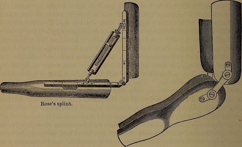
Rose's Splint on the left, Splints, Welch's Splints on the Right, amputation, Fractures, elbow joint
Contributed by Wikimedia Commons, (Public Domain)