Neuroanatomy, Superior Colliculus
- Article Author:
- Ryan Zubricky
- Article Editor:
- Joe M Das
- Updated:
- 7/31/2020 3:57:08 PM
- For CME on this topic:
- Neuroanatomy, Superior Colliculus CME
- PubMed Link:
- Neuroanatomy, Superior Colliculus
Introduction
The superior colliculus is a paired structure in the rostral midbrain that is involved in incorporating environmental stimuli and coordinating gaze shifts involving both eye and head movements. It is known as the optic tectum in other vertebrates and contains a topographic map of the contralateral visual field, as well as other inputs from somatosensory and auditory pathways.[1]
Structure and Function
The superior colliculus is on the posterior midbrain, rostral to the inferior colliculus, and caudal to the pineal gland. It has seven internal cell layers, divided into superficial, intermediate, and deep layers. The superficial layers consist of stratum zonale, stratum griseum superficiale, and stratum opticum. These layers respond to bilateral retinal inputs from contralateral visual stimuli and are purely sensory. The intermediate layers consist of the stratum griseum intermedium and the stratum album intermedium, while the remaining deep layers are the stratum griseum profundum and the stratum album profundum. Both of these deeper layers receive various sensory inputs from vision, auditory, and somatosensory pathways and are associated with motor function as well as sensory.
The superior colliculus has motor functions in the generation of eye movements. When head movement is restricted, the superior colliculi are involved in the saccadic motion of the eyes. Otherwise, the superior colliculi are involved in producing gaze shifts, which are initiated by saccades of the eyes, followed by a head movement with the eyes counter-rotating to stabilize. Thus, the superior colliculi are associated with both an oculomotor pathway and a head pathway responsible for producing saccades and movement in the neck muscles, respectively. Important to note is that while the superior colliculus encodes gaze shifts, its function does not include the specific movements needed to accomplish them. The superior colliculus, along with the inferior colliculus, is thought to function to orient the head and eyes toward stimuli that are seen and heard. Other nonmotor functions of the superior colliculus are less studied and include multimodal sensory processing and attention.[2][3][4]
Embryology
The mesencephalon serves as the embryonic precursor to the midbrain and thus the superior colliculus at both the primary and secondary vesicle stages of brain development at weeks 4 and 5, respectively. Expression of the specific genes Pax7 and engrailed have correlations with the formation and specialization of the superior colliculus, with the expression of Pax7 being essential for initiation of its creation as well as all stages of tectal development and engrailed expression being necessary for regulating cell migration during the formation of the tectal lamina.[5][6]
Blood Supply and Lymphatics
The superior colliculus receives its blood supply from both the collicular artery and the posteromedial choroidal artery. These are both proximal branches from the posterior cerebral artery. It is noteworthy that the superior cerebellar artery, which branches from the basilar artery, is sometimes attributed to supply the superior colliculus. Lymphatics of the brain were recently discovered, with one study detecting the presence of brain lymphatic endothelial cells over the optic tectum of zebrafish, which selectively uptake specific macromolecules.[7][8][9]
Nerves
The neural connections to and from the superior colliculus are complex and are still not entirely understood. As stated above, the superior colliculus receives visual information mainly from the retina via the optic nerve and tract, and less so from the visual cortex. The superior colliculus also receives auditory input from the inferior colliculus to coordinate a movement response. Additional somatosensory projections to the deep laminae allow the superior colliculus to respond to tactile stimuli. The superior colliculus also receives inputs from the prefrontal cortex involved in the regulation of attention and distractibility. These efferent fibers leave the internal capsule, follow a pedunculotegmental route to the lower midbrain, and then rise to the caudal superior colliculus. Outward tracts from the superior colliculus have been shown to project to the central gray matter overlying the rostral oculomotor nucleus as well as areas adjacent to the contralateral trochlear nucleus and contralateral abducens nucleus.[3][10][11]
Muscles
In the production of gaze shifts, various oculomotor and neck muscles ultimately receive stimulation by the activity of the superior colliculus. As stated above, the oculomotor, trochlear, and abducens nuclei all receive projections from the superior colliculus. Thus, the six extraocular muscles innervated by these nerves are all implicated in the motor actions of the superior colliculus. Additionally, the contralateral deep neck muscles are stimulated by the superior colliculus during a gaze shift. These muscles are ipsilateral to the visual field being stimulated and function to turn the head and eyes towards the stimulus.[2][10]
Surgical Considerations
Surgery of the superior colliculus should be approached with caution as the midbrain is an eloquent area associated with significant risks from surgical resection. Surgery is for tectal gliomas. However, complications include visual defects, Parinaud syndrome, mutism, and reduction to a vegetative state followed by death. Thus, surgical resection is often reserved based on the progression of the tumor. Treatment for tectal gliomas is still controversial and up for debate, yet with the advent of novel neurosurgical microsurgical techniques, this once inaccessible area is now approachable with increased safety.[12][13]
Clinical Significance
Direct damage to the superior colliculus has been tested in rhesus monkeys as well as rats and has resulted in clinical manifestations. Researchers found that the monkeys with damage to the superior colliculus to have visual deficits as well as impairments in gaze shifts. Rats with collicular damage displayed a lack of orienting reflex and no distraction when presented with visual or auditory stimuli when compared with normal or rats with visual cortex lesions. In humans, damage to the connection with the prefrontal cortex has shown to be associated with effects to attention. Lesions interrupting inhibitory inputs from the dorsolateral prefrontal cortex to the superior colliculus caused increased distractibility in one patient.
Tectal glioma, a rare low-grade tumor, is a condition of particular concern. It affects the superior and inferior colliculi as well as the cerebral aqueduct, resulting in increased intracranial pressure and long-term morbidities. It primarily affects a pediatric population with symptoms, including chronic headaches, visual deficits, and neurological impairment. Treatment traditionally involves shunting of the cerebrospinal fluid, and radiation therapy, chemotherapy, or possible surgery if the tumor continues to enlarge.[11][14][15][16][12]
(Click Image to Enlarge)
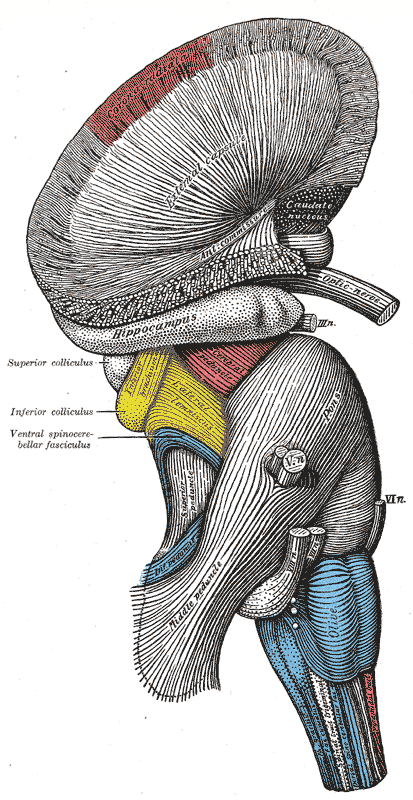
The Hind-brain or Rhombencephalon, Superficial dissection of brain-stem; Lateral view, External Capsule, Hippocampus, Superior colliculus, Inferior colliculus, Ventral Spinocerebellar fasciculus
Contributed by Gray's Anatomy Plates
(Click Image to Enlarge)
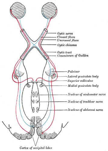
Central connections of the optic nerves and optic tracts, Crossed fibers, Uncrossed fibers, Optic chiasma, Optic tract, Commissure of Gudden, Pulvinar, Lateral geniculate body, Superior Colliculus, Medial Geniculate body, Nucleus of Oculomotor nerve, Nucleus of Trochlear nerve, Nucleus of abducens nerve
Contributed by Gray's Anatomy Plates