Syphilis
- Article Author:
- Maria Tudor
- Article Author:
- Ahmad Al Aboud
- Article Editor:
- William Gossman
- Updated:
- 8/10/2020 9:13:41 PM
- For CME on this topic:
- Syphilis CME
- PubMed Link:
- Syphilis
Introduction
Syphilis is a systemic bacterial infection caused by the spirochete Treponema pallidum. Due to its many protean clinical manifestations, it has been named the “great imitator and mimicker.” The origin of syphilis has been controversial and under great debate, and many theories have been postulated regarding this.
The pre-Columbian theory looked at findings on skeletal markers of syphilis before 1490. However, there is insufficient proof, as evidenced by the DNA and paleopathology findings, to support the existence of syphilis before 1492.
The Columbian and most accepted theory postulates that syphilis came from Europe in the 1490s when Columbus arrived in the New World (America). Syphilis spread when Christopher Columbus arrived in Naples (Italy). After Naples lost the battle to the French troops, this new disease spread across Europe.[1]
Syphilis remains a contemporary plague that continues to afflict millions of people worldwide.
The infection progresses through 4 stages and can affect many organ systems. Luckily, the organism treponema is still sensitive to penicillin.
Etiology
Treponema pallidum was identified as the agent that causes syphilis in 1905 by German scientists, and one year later, the test to diagnose this infection was developed. Its genome was sequenced in 1998. Treponema genus is a spiral-shaped bacteria with a rich outer phospholipid membrane that belongs to the spirochetal order. It has a slow metabolizing rate as it takes an average of 30 hours to multiply.
T. pallidum is the only agent that causes venereal disease. The other T. pallidum subspecies cause non-venereal disease that is transmitted via nonsexual contact: Treponema pertenue causes yaws, Treponema pallidum endemicum causes endemic syphilis, and Treponema carateum causes pinta. All the treponematoses have similar DNA but differ in their geographical distribution and pathogenesis.[2]
The only host for the organisms are humans, and there is no animal reservoir. Syphilis is considered a sexually transmitted disease, as most cases of syphilis are transmitted through vaginal, anogenital, and orogenital contact. The infection can rarely be acquired via nonsexual contact, such as skin-to-skin contact or via blood transfer (blood transfusion or needle sharing). Vertical transmission occurs transplacentally, resulting in congenital syphilis.
Epidemiology
According to the Center for Disease Contol and Prevention (CDC) statistics, there were 88,042 reported new diagnoses of syphilis in 2016. Out of all syphilis cases, 27,814 were primary and secondary syphilis. In 2016, most syphilis cases occurred among gay, bisexual, and other men who have sex with men. Men aged 20 to 29 years have the highest rates of primary and secondary syphilis.
From 2008 to 2012, rates of congenital syphilis declined but increased by 38% in 2012. During 2016, 628 cases of congenital syphilis were reported with rates 8.0-times and 3.9-times higher among infants born to black and Hispanic mothers compared to white mothers.
Syphilis is endemic in the developing world and is especially common among those who are poor and have limited access to health care.[3] Promiscuity plays an important role in disease transmission, as is more common among people with multiple partners.
Syphilis is an important synergistic infection for HIV acquisition and has been closely linked with HIV infection.
Pathophysiology
Treponema is a very tiny organism that is invisible on light microscopy. Thus, it is identified by its distinct spiral movements on darkfield microscopy. Outside the body, it does not survive for long.
The classic primary syphilis presentation is a solitary non-tender genital chancre in response to invasion by the T. pallidum. However, patients can have multiple non-genital chancres, such as digits, nipples, tonsils, oral mucosa. These lesions can occur at any site of direct contact with the infected lesion and are accompanied by tender or non-tender lymphadenopathy. Even without treatment, these primary lesions will go away without scarring. If untreated, primary syphilis can progress to secondary syphilis, which has many clinical and histopathological findings.
The clinical manifestations of secondary syphilis result from hematogenous dissemination of the infection and are protean: condyloma lata (papulosquamous eruption), hands and feet lesions, macular rash, diffuse lymphadenopathy, headache, myalgia, arthralgia, pharyngitis, hepatosplenomegaly, alopecia, and malaise. As a result, syphilis has been named the great imitator.
Both primary and secondary lesions resolve without treatment, and the patient enters either an early or latent phase in which no clinical manifestations are present. The infection can only be detected at this stage with serological testing. Some patients in this stage will progress to the tertiary stage, characterized by cardiovascular syphilis, neurosyphilis, and late benign syphilis.[3]
The incubation period is about 20 to 90 days. The organism does invade the CNS early, but symptoms appear late.
Histopathology
T. pallidum is a slowly metabolizing spirochetal bacterium, requiring an average of 30 hours to multiply and cannot be cultured on artificial media. Its outer membrane lacks lipopolysaccharides and has few surface-exposed proteins, making it difficult for the immune system to fight the infection. Because of this characteristic, T. pallidum is labeled as a stealth pathogen.
There are many histopathological features of syphilis, such as interstitial inflammation, endothelial swelling, irregular acanthosis, elongated rete ridges, a vacuolar pattern with lymphocytic infiltration. Silver staining can detect spirochetes anywhere from 30% to 70% but comes with a high rate of false-negative interpretation. Immunohistochemistry has a sensitivity of about 70% of accurately identifying the infection.[4]
History and Physical
Primary syphilis appears 10 to 90 days after exposure to the infection and comprises a painless, indurated ulcer (chancre) at the site of inoculation with the T. pallidum. HIV patients usually develop multiple chancres. These lesions resolve without treatment in 3-6 weeks. Regional lymphadenopathy is common and consists of rubbery lymph nodes.
Secondary syphilis appears 2 to 8 weeks after the disappearance of the chancre and has multiple systemic manifestations that can involve any system and body part. The cutaneous manifestations are also varied (condyloma lata, alopecia, mucous patches, palmar or truncal rash, papulosquamous rash) and because they contain a high load of spirochetes, these lesions are highly contagious.
Untreated primary or secondary syphilis is followed by an early latent phase (one year or less later on) or late latent phase (over 1 year) and is characterized by positive serologic tests, but negative clinical manifestations.
Tertiary syphilis is late symptomatic syphilis that can manifest months or years after the initial infection as cardiovascular syphilis (an aortic aneurysm, aortic valvulopathy), neurosyphilis (meningitis, hemiplegia, stroke, aphasia, seizures, tabes dorsalis), or gummatous syphilis (infiltration of any organ and its subsequent destruction).
Congenital syphilis results from transplacental transmission or contact with the infectious lesions during birth and can be acquired at any stage, causing stillbirth or neonate congenital infection. There are many presentations of congenital syphilis, including nasal cartilage destruction (saddle nose), frontal bossing (olympian brow), bowing of the tibia (saber shins), morbilliform rash, rhinitis (snuffles), sterile joint effusion (Clutton joints), peg-shaped upper central incisors (Hutchinson teeth). Many of the neonates born with congenital syphilis are asymptomatic at birth.[5] Early signs can manifest up to 48 months as rash, hepatosplenomegaly, fever, bulging fontanels, seizures, or cranial nerve palsies. Those untreated neonates enter a latent period. Routine screening is recommended at the first prenatal visit and during the third trimester and delivery in high-risk women.
Evaluation
Testing strategies for syphilis consist of dark-field microscopy and serological tests.
Dark-field examination by microscope allows for direct examination of spirochetes from the mucosal lesion and thus offers an immediate diagnosis.
The serological tests are classified as non-treponemal and treponemal. The non-treponemal tests (venereal disease research laboratory tests, rapid plasma reagin test) are screening tests that detect antibodies to cardiolipin in blood. The VDRL and RPR tests are only positive after the development of the primary chancre.
Positive non-treponemal tests are confirmed with treponemal tests (fluorescent treponemal antibody absorption assay, T. pallidum particle agglutination assay) that detects antibodies to the T. pallidum in blood. Syphilis is a reportable disease.
Patients with neurologic symptoms should undergo cerebrospinal (CSF) examination.
All patients with syphilis should be tested for other STDs. In addition, today, syphilis is routinely tested during the first trimester of pregnancy. If the testing is positive, benzathine penicillin G is administered.
Imaging studies depend on the organ involved. A chest x-ray may be the first clue to the presence of an aortic aneurysm. A CT scan can confirm this. An echocardiogram is needed to rule out aortic regurgitation.
Reverse sequence screening is an increasingly used algorithm across US laboratories that use treponemal tests as the initial screening to identify those patients with treated, untreated, or incompletely treated syphilis.[6] Because of a lack of validation of the reverse algorithm, higher rates of false-positive results can be seen, leading to difficulty in interpreting these tests and the need for second confirmatory treponemal tests.
Treatment / Management
Treatment depends on the disease stage.
Primary, secondary, or early latent syphilis is treated with a single dose of intramuscular (IM) penicillin G benzathine 2.4 million units. Alternative therapies include doxycycline 100 mg orally (PO) twice daily for 14 days or ceftriaxone 1 to 2 gm IM or intravenously (IV) daily for 10 to 14 days or tetracycline 100 mg PO 4 times for 14 days.
Late latent syphilis is treated with IM penicillin G benzathine 2.4 million units once weekly for 3 weeks. Alternative therapies include doxycycline 100 mg PO twice daily for 28 days or tetracycline 100 mg PO four times daily for 28 days.
Tertiary syphilis is treated with IM penicillin G benzathine 2.4 million units once weekly for 3 weeks.
Neurosyphilis is treated IV penicillin G aqueous 18-24 million units daily for 10 to 14 days.
Patients with a high titer of secondary syphilis can develop Jarisch-Herxheimer reaction, which is an immune-mediated self-limited reaction that occurs within 2 to 24 hours of treatment and is characterized by high fever, headache, myalgias, rash.
Patients need to be followed post-treatment at 3, 6, 9, 12, and 24 months with serial non-treponemal tests. A 4-fold decline in these tests indicates successful treatment.[7][8]
Jarisch Herxheimer Reaction
Following treatment with penicillin, the dying organisms often release inflammatory cytokines that lead to the Jarisch Herxheimer reaction. The symptoms include headache, muscle pain, fever, tachycardia, and malaise. The reaction usually appears within 24 hours of starting treatment. The treatment is supportive. Pregnant women who develop this reaction need to be observed closely as it can lead to obstetric complications.
Preventing Syphilis
If a person has had sex with an infected individual, benzathine penicillin is administered.
Differential Diagnosis
- Genital herpes
- Behcet syndrome
- Mononucleosis
- Contact or atopic dermatitis
- Lymphoma
- Viral exanthema
- Pityriasis rosea
- Erythema multiforme
- Rocky Mountain spotted fever[3]
Pertinent Studies and Ongoing Trials
WHO guidelines
- Benzathine penicillin administered intramuscularly is the treatment of choice
- Procaine penicillin is the 2nd choice and administered for 10 to 14 days intramuscularly
- If penicillin cannot be used, doxycycline, azithromycin or ceftriaxone are other options
- Doxycycline is preferred as it is cheap and easy to administer. But it is not recommended in children or pregnant women
- Azithromycin does not cross the placenta and hence the infant has to be treated after delivery
Prognosis
The prognosis of syphilis depends on the stage and extent of organ involvement. If left untreated, the organism has significant morbidity and mortality. Patients usually develop cardiovascular and CNS syphilis, which are fatal. Congenital syphilis is associated with spontaneous abortions, stillbirth, and fulminant pulmonary hemorrhage in neonates. Without treatment during pregnancy, syphilis is almost always passed on to the fetus.
Complications
Untreated syphilis infection can lead to irreversible neurological and cardiovascular complications. Depending on the stage, neurosyphilis can manifest as meningitis, stroke, cranial nerve palsies during early neurosyphilis or tabes dorsalis, dementia, general paresis during late neurosyphilis. Cardiovascular syphilis is also a result of tertiary syphilis and can manifest as aortitis, aortic regurgitation, carotid ostial stenosis, or granulomatosis lesions (gummas) in various body organs.
Untreated syphilis affects the course of HIV infection with higher virus replication and lowers CD4 counts and a faster rate of progression to late syphilis.[8] Primary and secondary syphilis during pregnancy lead to neonatal infection and adverse pregnancy outcomes if not timely treated.
Consultations
It is recommended for the primary physician to consult with an infectious disease specialist regarding confirmation of disease, treatment, and follow-up. Pediatric infectious disease specialists are best suited in the management of congenital syphilis.
Pearls and Other Issues
Syphilis can cause a gamut of systemic manifestations, and for this reason, it has been called the “great mimicker.” Despite its discovery centuries ago, it continues to be a major public health problem. Because of its varied manifestations, diagnosis can be challenging. Physicians need to maintain an increased suspicion index for screening high-risk populations, such, men who have sex with men, pregnant women with syphilis, HIV-infected patients. Penicillin remains the main treatment based on the stage of infection and whether CNS involvement occurs. Treated patients need to be followed-up with non-treponemal antibody titers to evaluate treatment response.
Enhancing Healthcare Team Outcomes
Once the diagnosis of syphilis has been made, the management is with an interprofessional team since the infection can affect almost every organ in the body. These patients need close follow up by the cardiologist, neurologist, dermatologist, internist, ophthalmologist, obstetrician, and the infectious disease expert. The patient must be followed by the infectious disease nurse to ensure that the treatment is working and the patient is compliant with therapy. The patient's partner has to be investigated and treated if positive. If the patient with syphilis is pregnant, close follow up with an obstetrician is highly recommended.[9][10]
Patient education is vital, and the public health nurse should counsel patients against the use of IV drugs or use clean needles. Today, there are needle exchange programs in almost every city. In addition, the nurse has to educate the public on safe sex practices and the importance of regular screening for sexually transmitted diseases. The use of barrier protection (condom) is highly recommended. The pharmacist should educate the patient that there are effective treatments for STDs, and the earlier the condition is treated, the better the outcomes. Only through close collaboration with an interprofessional team can the morbidity of infections like syphilis be lowered.
The outlook for most patients who are compliant with treatment is good, but those who delay or fail to comply with treatment can develop life-threatening complications.[11][12]
(Click Image to Enlarge)
Syphilis
Contributed by DermNetNZ
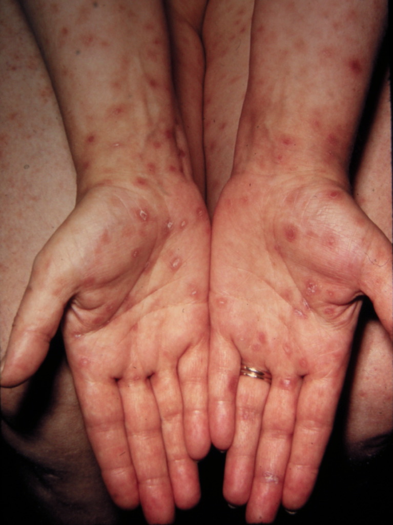
(Click Image to Enlarge)

This photograph shows a close-up view of keratotic lesions on the palms of this patient’s hands due to a secondary syphilitic infection. Syphilis is a complex sexually transmitted disease (STD) caused by the bacterium Treponema pallidum. It has often been called "the great imitator" because so many of the signs and symptoms are indistinguishable from those of other diseases.
Contributed by Wikimedia Commons, Robert Sumpter (CDC) (Public Domain)
(Click Image to Enlarge)

Fundoscopic image, effect of late neuro-ocular syphilis on the optic disk and retina, Pathology, Severe optic nerve atrophy, chorioretinitis, inflammation of the choroidal and neural layers of the retina
Contributed by Susan Lindsley, The Centers for Disease Control and Prevention (CDC)
(Click Image to Enlarge)

Syphilis gummas lesion
Contributed by The Centers for Disease Control and Prevention (CDC)
(Click Image to Enlarge)

Primary Syphilis Chancre
Contributed by Dr. Shyam Verma, MBBS, DVD, FRCP, FAAD, Vadodara, India