Tinea Corporis
- Article Author:
- Garrett Yee
- Article Editor:
- Ahmad Al Aboud
- Updated:
- 8/14/2020 8:55:09 PM
- For CME on this topic:
- Tinea Corporis CME
- PubMed Link:
- Tinea Corporis
Introduction
Tinea corporis is a superficial fungal skin infection of the body caused by dermatophytes. Tinea corporis is present worldwide. It is defined explicitly by the location of the lesions that may involve the trunk, neck, arms, and legs. Alternative names exist for dermatophyte infections that affect the other areas of the body. These include the scalp (tinea capitis), the face (tinea faciei), hands (tinea manuum), the groin (tinea cruris), and feet (tinea pedis).
Etiology
The dermatophyte's ability to attach to the keratinized tissue of skin forms the basis for the dermatophytoses (superficial fungal skin infections).[1] The dermatophytes causing tinea corporis belong to genera Trichophyton, Epidermophyton, and Microsporum. Trichophyton rubrum is the most common species to cause dermatophyte infections in the last 70 years. T. Rubrum accounts for 80 to 90% of the strains.[2] Other common isolates include Trichophyton mentagrophytes and Microsporum audouinii. Infection typically occurs with direct contact with the skin from the soil, animals, or the skin of other humans.
In some individual cases, the most common etiologic species is dependent on the method of transmission. Tinea corporis secondary to Trichophyton tonsurans (T. tonsurans) commonly results from direct contact with a patient with tinea capitis. In tinea capitis, T. tonsurans is the most common causative agent in the United States and the United Kingdom.[3][4] T. tonsurans is also a typical finding in cases of tinea corporis gladiatorum.[5] Tinea corporis gladiatorum can present in athletes with extensive direct skin-to-skin contact (classically wrestlers). Patients with tinea corporis with close contact with cats or dogs commonly are infected with Microsporum canis.
Epidemiology
Tinea corporis is exceedingly common worldwide. Dermatophytes are the most prevalent agents of superficial fungal infections. Excessive heat, high relative humidity, and fitted clothing have correlations to more severe and frequent disease.[6] Specific populations can also be more predisposed to tinea corporis; for example, children. Tinea capitis and tinea corporis are the most common dermatophytic infections in prepubertal children.[7] Children are also more likely to contract zoophilic infections. Zoophilic infections get transmitted via contact with animals such as cats and dogs. Another vulnerable population includes patients with compromised immune systems. The immunocompromised patients also have an increased prevalence in developing Majocchi granuloma, a type of tinea corporis folliculitis that invades the deep dermal layers in contrast to the more superficial traditional tinea corporis.[8]
Pathophysiology
All people do not have equal susceptibility to fungal infection, and there are familial and genetic predispositions possibly mediated by specific defects in innate and adaptive immunity. Patients with low defensin beta 4 may demonstrate a predisposition to all dermatophytes. Some other predisposing factors include underlying diseases such as diabetes mellitus, lymphomas, immunocompromised status, Cushing syndrome, excess sweating, or old age. The currently held view is that a cell-mediated immune response is responsible for the control of dermatophytosis.
Histopathology
Microscopic examination takes place after administration of 10% to 20% potassium hydroxide (KOH) solution to the skin scrapings. Gentle heating, in addition to the KOH preparation, serves to dissolve the keratin and emphasize the dermatophyte itself. Microscopic visualization reveals septate and branching long, narrow hyphae without constriction.
History and Physical
Patients commonly present with an itchy, red rash. These typically present on the exposed skin of the neck, trunk, and/or extremities.
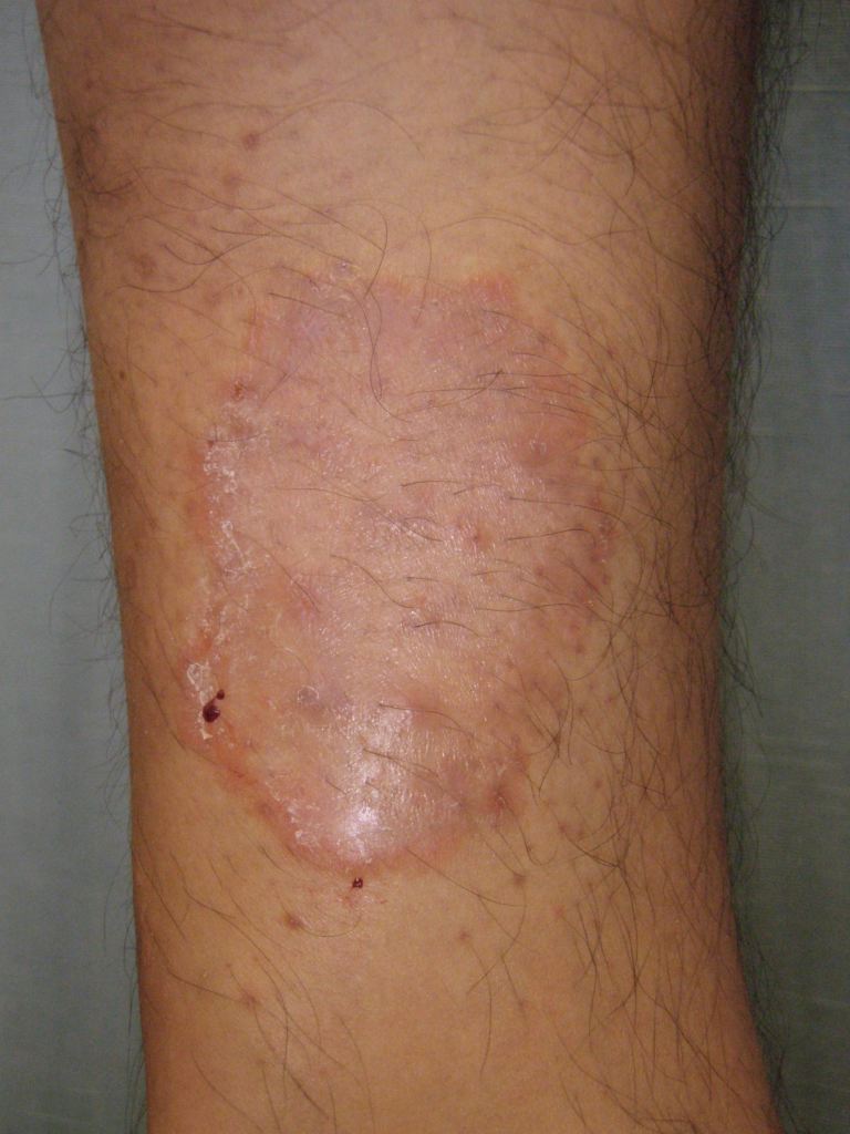
On physical exam, single or multiple lesions are usually circular or ovoid in appearance with patches and plaques. These annular lesions demonstrate sharp marginations with a raised erythematous scaly edge which may contain vesicles. The degree of inflammation is variable.
The lesions advance centrifugally from a core, leaving a central clearing and mild residual scaling; this appears as a "ring" shape giving rise to the term "ringworm."
Evaluation
The diagnosis of tinea corporis is usually clinically based on a thorough history and physical examination. However, testing can be done to confirm the diagnosis. Skin scrapings examined under a microscope with a potassium hydroxide (KOH) preparation will reveal septate and branching long narrow hyphae. However, up to 15% of cases may yield false negatives when only using KOH preparations for diagnosis.[9]
Therefore another method for confirmation is a fungal culture. Fungal cultures are possible but take time for definitive identification. Cultures may begin to see growth in about five days but may take up to four weeks in certain species. Therefore at least four weeks are needed to deem a sample as "no growth." The most common isolation medium used for fungal cultures is a Sabourad dextrose agar (1% glucose, 4% mycological peptone agar, water). Identification is by examining the morphology, pigmentation, and surface topography of the culture.
Treatment / Management
The treatment of dermatophyte infections usually involves the use of topical or oral preparations.
Localized tinea corporis commonly responds to topical therapy typically applied once or twice daily, usually for two to three weeks. However, the endpoint of therapy is a clinical resolution of the symptoms. In general, nystatin topical is not effective for tinea corporis.
Suggested topical regimens include one of the following:
- Clotrimazole: 1% cream/ointment/solution applied topically twice daily
- Ketoconazole: 2% cream/shampoo/gel/foam applied once daily
- Miconazole: 2% cream/ointment/solution/lotion/powder applied twice daily
- Naftifine: 1% cream, applied once daily or 1% or 2% gel twice daily
- Terbinafine: 1% cream/gel/spray solution once or twice daily
Oral therapy is necessary in more widespread infections or cases that have failed topical treatment. Oral terbinafine or itraconazole is usually the preferred first-line treatments and is expected to clear the condition in about 2 to 3 weeks.
Suggested oral regimens include one of the following (for adults):
- Terbinafine: 250 mg orally once daily for two weeks
- Itraconazole: 100 mg once daily for 2 weeks or 200 mg once daily for one week; give capsules with food
- Fluconazole: 150 to 200 mg once weekly or 50 to 100 mg/day for 2 to 4 weeks
- Griseofulvin: 500 to 1000 mg once daily for 2 to 4 weeks
Differential Diagnosis
Diseases that are in the differential diagnosis may mimic the appearance of tinea corporis. These also typically present with annular lesions. Cases that are refractory to antifungal treatment or have a negative potassium hydroxide microscopic examination should warrant further investigation. Also, the clinician must rule out other, more serious conditions if there is a severe disease such as extensive skin involvement.
Other common diseases that may present similarly include nummular eczema, erythema annulare centrifugum, tinea versicolor, cutaneous candidiasis, subacute cutaneous lupus erythematosus, pityriasis rosea, contact dermatitis, atopic dermatitis, seborrheic dermatitis, psoriasis.
Some of the severe diseases that must be ruled out are secondary syphilis, mycosis fungoides, or parapsoriasis.
Prognosis
Prognosis is usually good with proper treatment and patient compliance.
Complications
Complications are uncommon in dermatophytic infections.
One such complication includes Majocchi granuloma, is a rare condition in which the dermatophyte invades via a follicle and advances deeper into the dermis or subcutaneous tissue. Minor skin trauma such as shaving can predispose patients to Majocchi granuloma. Lesions involve the hair follicles and appear as erythematous nodules or papules. These may even progress to abscesses. Oral antifungals such as terbinafine 250 mg once daily for 2 to 4 weeks are the recommended therapy in cases of Majocchi granuloma.
Deterrence and Patient Education
Education is paramount is preventing tinea corporis. Patients may be encouraged to wear light and loose fitting clothing. Also keeping the skin clean and dry will help prevent the development of tinea corporis.
Also, upon initiation of topical antifungal treatment, compliance needs to be encouraged; however, the results are typically not immediate. The patient may need reminders that even with proper treatment, it may take weeks to begin seeing the resolution of symptoms.
Pearls and Other Issues
Hepatitis is a proven complication with ketoconazole, so baseline liver panel is necessary before initiating oral antifungals, especially the azole group. Terbinafine is associated with a lupus-like reaction, so it should be given cautiously in patients with systemic lupus erythematosus. Additionally, allergic contact dermatitis to topicals is possible but rare, which means irritant effects may occur.
Enhancing Healthcare Team Outcomes
Tinea corporis can be diagnosed clinically based on history and exam. If there is an atypical appearance, further testing such as a KOH test or fungal culture should be the diagnostic test plan. [Level III]
After diagnosing tinea corporis, the standard treatment is with topical antifungals. Due to the possibility of side effects and adverse reactions from systemic therapy, topical treatment is the usual recommendation over systemic therapy. [Level I]
Tinea corporis requires an interprofessional team approach, including physicians, specialists, specialty-trained nurses, and pharmacists, all collaborating across disciplines to achieve optimal patient results. [Level V] In most cases, the clinician (physician, NP, PA) will diagnose and prescribe treatment. Pharmacists can verify agent antifungal coverage and dosing, and report back to the nurse or clinician if they have any concerns. Pharmacists and nurses can both ensure that the patient has had baseline LFTs and report back to the prescriber if not. The pharmacist should also verify the patient's current regiment has no other drugs that could contribute to hepatotoxicity because of potential add-on effects of many oral antifungal agents. If present, the pharmacist should alert the nurse or prescriber so they can arrange for appropriate changes to the therapeutic regimen. Pharmacists can also check for drug-drug interactions, as these are common with azole antifungals. Nurses and pharmacists can both verify patient compliance and counsel patients on their medications or the dosing/administration of the same, and report any issues back to the prescribing clinician, who can make changes to the patient's drug regimen based on patient needs.

