Urticaria Pigmentosa
- Article Author:
- Angela Macri
- Article Editor:
- Christopher Cook
- Updated:
- 8/10/2020 4:18:19 PM
- For CME on this topic:
- Urticaria Pigmentosa CME
- PubMed Link:
- Urticaria Pigmentosa
Introduction
Mastocytosis is a disorder characterized by mast cell accumulation, commonly in the skin, bone marrow, gastrointestinal (GI) tract, liver, spleen, and lymphatic tissues. The World Health Organization (WHO) divides cutaneous mastocytosis into three main presentations. The first has solitary or few (less than or equal to 3) lesions called “mastocytomas.” The second involves multiple lesions ranging from less than 10 to greater than 100 which is referred to as “urticaria pigmentosa” (UP). The last presentation involves diffuse cutaneous involvement. Urticaria pigmentosa is the most common cutaneous mastocytosis in children, and it can form in adults as well. It is thought to be a benign, self-resolving condition that remits in adolescence. Unlike adult forms of mastocytosis, there is rarely any internal organ involvement in UP.[1]
Etiology
UP is caused by several activating mutations in the KIT gene. When exposed to certain triggers, mast cells release mediators that cause the symptoms of mastocytosis. The mediators that are released are histamine, eicosanoids, prostaglandins, leukotrienes, heparin, proteases, and cytokines. Triggers include certain foods, exercise, heat, Hymenoptera and venomous stings, local trauma to skin lesions, alcohol, narcotics, salicylates and other nonsteroidal anti-inflammatory drugs (NSAIDs), polymyxin B, and anticholinergic medications. Some systemic anesthetic agents may induce anaphylaxis.[2]
Epidemiology
Mastocytosis can present at birth or develop any time into late adulthood. It occurs in all races and has no gender preferences. Although most patients do not have a family history of mastocytosis, familial cases have been reported. Cutaneous mastocytomas occur in 15% to 50% of patients, urticaria pigmentosa occurs in 45% to 75%, and diffuse cutaneous involvement occurs in less than 5% to 10%.
Pathophysiology
Mast cell precursors express CD34, the tyrosine kinase receptor KIT (CD117), and IgG receptors (Fc gamma RII). KIT can be activated by its ligand (stem cell factor, SCF) which induces mast cell growth and maturation and prevents apoptosis. Bone marrow stromal cells, keratinocytes, endothelial cells, fibroblasts, and reproductive Sertoli, and granulosa cells produce SCF. The pathogenesis of mastocytosis is due to several mutations in KIT. The most common is an activating mutation in codon 816 where there is a substitution of the amino acid aspartic acid (D) with valine (V, i.e., D816V) or another amino acid. These amino acids result in constitutive ligand-independent activation of the receptor. Other additional pathogenic factors exist that still need more research.[3][4]
Histopathology
Diagnosis of UP can be made clinically. However, a definitive diagnosis requires a skin biopsy. The hallmark finding histologically for UP is having an increased amount of mast cells in the dermis. UP skin lesions can have up to a 40-fold higher mast cell count than normal skin. Mast cells have a rounded or cuboidal appearance. Stains that help identify mast cells include toluidine blue, Leder, Giemsa, tryptase, and CD117 (KIT). Eosinophils are commonly found in the dermis and hyperpigmentation of the basal layer can also be found. Molecular studies for KIT mutations can be performed on biopsy specimens.
Biopsy specimens from normal skin in patients with mastocytosis will not show an increased amount of mast cells. Therefore, if there are no skin lesions and systemic mastocytosis is suspected, a bone marrow biopsy, or biopsy from the GI tract may be needed. A total serum tryptase level can help in identifying the extent of mast cell disease. A tryptase level greater than 20 ng/ml represents one of the minor criteria for systemic mastocytosis.[5][1]
History and Physical
Urticaria pigmentosa has small, monomorphic tan to brown macules or papules distributed mostly on the trunk and classically spares the central face, palms, and soles. It is similar to the lesions typically observed in adults. Lesions can be few or numerous and are usually about 1 to 2 cm in size however they can be larger. Darier’s sign is the formation of a wheal upon stroking or rubbing skin lesions and suggests the diagnosis of cutaneous mastocytosis. The wheal forms due to the release of mast cell mediators which can contribute to the systemic symptoms that may occur after stroking the lesion. Some lesions may blister after stroking the lesion. Darier’s sign is more pronounced in children than it is in adults due to an increased density of mast cells in lesions of children.
Systemic symptoms can occur such as pruritus, flushing, abdominal pain, diarrhea, palpitations, dizziness, and syncope. Twenty-five percent of patients with UP experience gastrointestinal symptoms. Complaints of fever, night sweats, bone pain, epigastric distress, malaise, weight loss, and problems with cognitive disorganization often signal the presence of extracutaneous disease. Death can even occur from extensive mast cell mediator release.[6][2]
Evaluation
Start by obtaining a thorough history and physical. If the patient complains of GI symptoms, a barium study or endoscopy may be needed. If a patient complains of bone pain or fractures a radiographic skeletal survey or bone scan may be needed. On physical exam look for lymphadenopathy and hepatosplenomegaly. If you suspect an abnormality, order a liver ultrasound or CT scan to investigate. If there are abnormal findings in any of the tests, a biopsy of the bone marrow should be considered. A complete blood count (CBC), serum tryptase level, LFTs, and KIT gene analysis can be ordered. However, it is considered by some to be optional in the pediatric population. If there are any abnormalities, a bone marrow biopsy should be considered. Bone marrow involvement is not very common in children with cutaneous mastocytosis, as opposed to adults, and a bone marrow biopsy is not recommended for them. Although this paper focuses on urticaria pigmentosa, it is important to learn the criteria for systemic mastocytosis in case a patient progresses. [7]
The WHO criterion for the diagnosis of systemic mastocytosis states that you need 1 major and 1 minor criteria or 3 minor criteria. Criteria are listed below:
Major criteria
- Having multifocal, dense infiltrates of mast cells (aggregates of greater than or equal to 15 mast cells) in bone marrow or extracutaneous tissues
Minor criteria
- Greater than 25% of mast cells in bone marrow samples or extracutaneous tissues are spindle-shaped or otherwise atypical
- Expression of CD25 and/or CD2 by extracutaneous mast cells (often determined by bone marrow flow cytometry)
- Presence of activating KIT codon 816 mutation in blood, bone marrow, or extracutaneous tissues
- Serum total tryptase level greater than 20 ng/ml (exception would be an associated clonal myeloid disorder, then this is not valid)
The criterion for diagnosing cutaneous mastocytosis is not well defined. Cutaneous mastocytosis is usually diagnosed by visual evaluation of typical skin lesions, particularly in children. However, there is a stepwise approach to diagnose mastocytosis in the skin. Must have 1 major and 1 minor criterion.
Major criteria
- Must have the typical skin lesions.
Minor criteria
- Histology (monomorphic mast cell infiltrate with aggregates of greater than 15 mast cells per cluster or scattered mast cells with greater than 20 per high microscopic power field)
- Molecular criterion (detection of a KIT mutation at codon 816 in the affected skin)[5]
Treatment / Management
Treatment is symptomatic for patients with cutaneous mastocytosis. Topical medications include calcineurin inhibitors and corticosteroids. Systemic therapies such as oral antihistamines (the mainstay of treatment), oral cromolyn sodium (used specifically for GI symptoms), oral corticosteroids, omalizumab, oral PUVA, narrowband ultraviolet B, and UVA1 can be used. A pre-measured epinephrine pen with auto-injector should be given to all UP patients. Patients should be educated to avoid possible triggers. [8]
Differential Diagnosis
Bullous impetigo, urticaria, juvenile xanthogranuloma, arthropod stings, auto-immune bullous diseases are differential diagnoses.
Prognosis
The prognosis for childhood mastocytosis is excellent with 50% to 70% of patients remitting before adolescence.
Enhancing Healthcare Team Outcomes
Urticaria pigmentosa is generally benign. an interprofessional team of clinicians and specialty trained nurses should educate the patient and family and provide support as needed. It will generally resolve in adolescents. [Level V]
(Click Image to Enlarge)
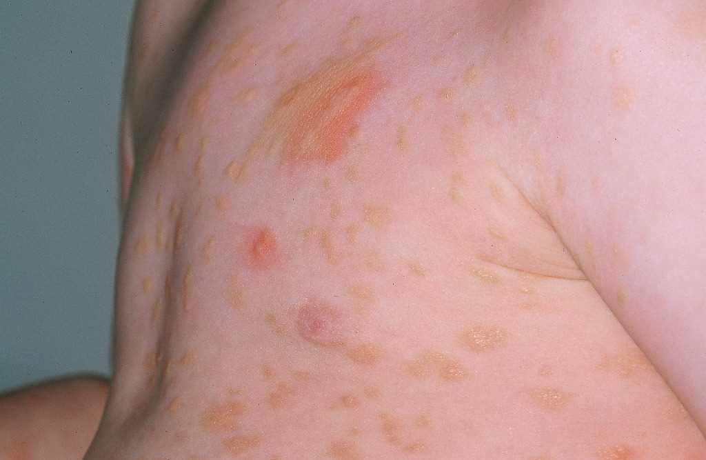
Urticaria Pigmentosa
Contributed by DermNetNZ