Anatomy, Back, Vertebral Canal
- Article Author:
- Tucker Peabody
- Article Editor:
- Joe M Das
- Updated:
- 10/28/2020 1:01:58 AM
- For CME on this topic:
- Anatomy, Back, Vertebral Canal CME
- PubMed Link:
- Anatomy, Back, Vertebral Canal
Introduction
The vertebral canal, otherwise known as the vertebral cavity or spinal cavity, is an anatomical space formed by the vertebral column that stores an integral portion of the central nervous system: the spinal cord and the spinal nerve roots branching off the spinal cord bilaterally. The vertebral canal is an anatomically sterile space that is considered a subdivision of the dorsal body cavity, which contains the cranial cavity and the vertebral cavity/canal. The vital function of the vertebral canal is serving as a protective space for the spinal cord to traverse the length of the spinal column allowing proper innervation to the entire human body.
Structure and Function
The vertebral canal is composed of the vertebral foramen located in the cervical, thoracic, and lumbar vertebrae. The vertebral or spinal canal typically ends at the level of the L2 vertebra, where the spinal cord gives off multiple branching spinal nerve and nerve rootlets known as the cauda equina. These spinal nerves traverse at the levels of the second through the fifth lumbar vertebral levels.
The vertebral foramen forms by the posterior portion of the vertebral body, the pedicles, and lamina laterally on either side and the connection of the lamina at the transverse process. The only exception to this anatomical formation is that the atlas and axis (C1 and C2 vertebra) lack a vertebral body whereas the atlas is comprised of an anterior and posterior arch, and the axis is made up of the odontoid process that projects into the vertebral foramen of the atlas. Two crucial structural ligaments surround the vertebral canal. These ligaments include the ligamentum flavum and the posterior longitudinal ligament. The ligamentum flavum is a ligament that traverses the entire spinal column, and it connects the lamina of each vertebra. The posterior longitudinal ligament runs along the posterior portion of the vertebral body, which is the anterior border of the vertebral canal.[1]
The spinal canal diameter varies with each section of a vertebra, including the cervical, thoracic, and lumbar regions. The average diameter of the cervical vertebra is 17mm. The thoracic vertebral canal varies with each vertebra moving in a cranial-caudal direction. Starting at the T1 vertebral level, the spinal canal diameter is 16 mm on average, and the diameter begins to decrease at the T2 level to 14-15 mm. The T4 vertebra has the smallest diameter moving caudally, the spinal canal from T3-T11 canal diameter remains relatively stable at 15 mm. At the T12 vertebra, the spinal canal diameter increases to around 18 mm on average. The lumbar vertebral group, compared to the cervical and thoracic regions, have a larger spinal canal diameter on average, with the L5 diameter being the largest of the lumbar vertebra at around 17.5 mm.
The spinal cord is the major structure contained within the vertebral canal. At each vertebral level, the spinal cord gives off projections bilaterally that project out of the vertebral canal. These are the spinal nerves that contain nerve fibers from the dorsal and ventral roots of the spinal cord. The dorsal root nerves carry sensory innervation traveling back to the spinal cord from the rest of the body, and the ventral roots contain motor innervation exiting from the spinal cord to travel throughout the human body.[2]
The main, and most important, the function of the vertebral canal is to provide a patent and protective cavity for the spinal cord to traverse from the cranium down to the sacrum. Not only does the vertebral column provide structure and support to the human body, but its canal provides physical protection for the spinal cord.
Embryology
Embryological formation of the vertebral column secondarily gives rise to the vertebral canal. During gastrulation, there are three germ layers, consisting of the mesoderm, endoderm, and ectoderm, the paraxial portion of the mesoderm is what will differentiate into forty-two pairs of segments, somites, that develop in a cephalocaudal direction during embryological development. The somites will further develop into dermatomyotomes and sclerotomes. The sclerotomes are what will later develop into the bony structure of the human spine.[3]
The ossification and development of the individual vertebra that comprises the vertebral column allows for the vertebra to contain a foramen through which the spinal cord traverses. The ossification of the vertebra is very specific, and it includes a total of eight different ossification centers: three primary centers coupled with five secondary centers. The primary centers include the bilateral neural processes and the centrum of the vertebra, and the secondary ossification centers include the transverse process tips, the tip of the spinous process, and the superior and the inferior surfaces of the vertebral body.[3]
Blood Supply and Lymphatics
The spinal cord that travels through the vertebral canal primarily receives its blood supply from two sources: anterior spinal artery and the posterior spinal artery. As these two arteries traverse the length of the spinal column, they give off branching arteries bilaterally at each vertebral level. These branching arteries are the segmental spinal arteries, and they enter into the vertebral canal through the intervertebral foramen.
The artery of Adamkiewicz, otherwise known as the great anterior radiculomedullary artery, is a perfusing branch of the radicular anterior artery that arises from the posterior intercostal artery. There are multiple anatomic variants of this artery and what vertebral level it joins with the anterior spinal artery.[4]
Clinical Significance
VACTERL syndrome is a multi-factorial condition that affects the embryological development of multiple organ systems. One of those systems is the skeletal system and, more specifically, the vertebral column. This condition can lead to skeletal malformations such as missing, fused, or even extra vertebra present in the spine that can compromise the spinal cord due to variation in size of the vertebral canal in which the spinal cord resides.[5]
Spondylolisthesis is the condition where a vertebral body will slip anteriorly relative to the inferior vertebral body. This condition can arise from a congenital malformation of the lumbosacral joint L5-S1, or it can more commonly occur in people you repetitively hyperextend and rotate their movements—this why spondylolisthesis most commonly presents in the gymnastics, swimming, diving, and weightlifting populations. If there is severe anterior slippage, typically less than 50% displacement of the superior vertebra concerning its inferior vertebra, there can be neurological deficits present due to compression of the spinal cord. The slippage causes a reduction in the vertebral canal space resulting in a compressed spinal cord. Spondylolisthesis can also be associated with spondylolysis, which is a bilateral fracture of the pars interarticularis portion of the vertebra. When this occurs, the stabilization of the vertebral column is compromised, and the vertebral body is now allowed to slip anteriorly.[6][7]
Disc herniation is when the nucleus pulposus portion of the intervertebral disc herniates through the weakens annular fibrosis fibers of the outer disc, causing compression of the spinal cord and compression of the spinal nerve roots. The nucleus pulposus herniates into the vertebral canal when the annular fibers become weakened, which is associated with degenerative disc disease.[8] There are multiple types of disc herniations, including protrusion, extrusion, and sequestration. One way to differentiate between a disc protrusion and extrusion is the ratio of the length of the apex of the herniated disc compared to the length of the base of the herniated disc portion. Disc protrusion is when the base length is greater than the length of herniated disc apex, and for extrusion, the length of the herniated apex is greater than the base of the herniated portion of the disc. Disc sequestration occurs when a herniated portion of the nucleus pulposus portion of an intervertebral disc fragment from the disc and becomes a free-floating segment within the spinal canal, allowing it to migrate caudally or cranially.[9][10]
Spinal stenosis occurs when there is a narrowing of the spinal canal resulting in spinal cord compression. There are multiple etiologies of spinal stenosis, including degenerative bone diseases such as osteoarthritis, rheumatoid arthritis, and even Paget disease, which can lead to bony overgrowth due to dysregulation of osteoblast and osteoclast activity. Neurovascular compression, due to the stenotic process, can lead to neurological pain symptoms that, depending on the severity and patient tolerance, can be very debilitating.[11][12]
(Click Image to Enlarge)
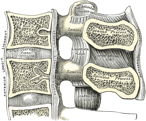
Vertebrae, Lumbar, Ligament, Medial Sagittal Section, Anterior Longitudinal Ligament, Inter vertebral Fibrocartilage, Posterior Longitudinal Ligament, Lamina, Ligamenta Flava, Pedicle, Spinous Process, Interspinal Ligament, Capsular ligament, Supraspinal Ligament,
Contributed by Gray's Anatomy Plates
(Click Image to Enlarge)

The Mediastinum, Transverse section through the upper margin of the second thoracic vertebra
Contributed by Gray's Anatomy Plates
(Click Image to Enlarge)
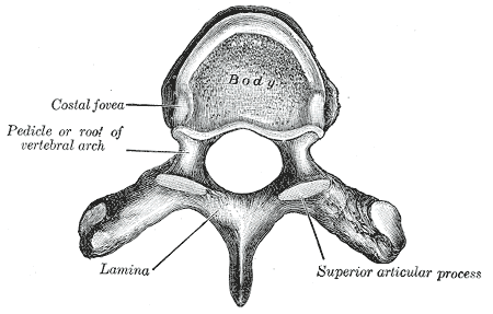
General Characteristics of Vertebrae, Costal Fovea, Lamina, Superior Articular process
Contributed By Gray's Anatomy Plates