Anatomy, Bony Pelvis and Lower Limb, Femur
- Article Author:
- Amy Chang
- Article Author:
- Grant Breeland
- Article Editor:
- John Hubbard
- Updated:
- 8/22/2020 7:13:48 PM
- For CME on this topic:
- Anatomy, Bony Pelvis and Lower Limb, Femur CME
- PubMed Link:
- Anatomy, Bony Pelvis and Lower Limb, Femur
Introduction
The femur is the longest, heaviest, and strongest bone in the human body. At the proximal end, the pyramid-shaped neck attaches the spherical head at the apex and the cylindrical shaft at the base. There are also 2 prominent bony protrusions, the greater trochanter and lesser trochanter, that attach to muscles that move the hip and knee. The angle between the neck and shaft, also known as the inclination angle is about 128 degrees in the average adult. However, the inclination angle decreases with age.[1][2]
The hip is a ball-in-socket joint that is composed of the acetabulum of the pelvis encompassing the femoral head. The head is pointed in a medial, superior, and slightly anterior direction. Ligamentum teres femoris connects the acetabulum to the fovea capitis femoris, which is a pit on the head.
The shaft has a mild anterior arch. At the distal femur, the shaft flares out in a cone-shaped manner onto a cuboidal base made up of the medial and lateral condyle. Medial and lateral condyle join the femur to the tibia, forming the knee joint.
Both hips and knees are synovial joints that are covered by cartilage to reduce friction and optimize the range of motion. Bony features are landmarks for measuring axis along the femur.[3][4]
Structure and Function
The main function of the femur is weight bearing and gait stability. The upper body’s weight sits on the 2 femoral heads. The capsular ligament is a strong thick sheath that wraps around the acetabulum periosteum and proximal femur.[5] It holds the femoral head within the acetabulum of the pelvis. The capsular ligament limits internal rotation but allows for external rotation.[6][7]
The knee is a hinge joint between the distal femur and proximal tibia. Medial and lateral meniscus stabilize and cushion the tibiofemoral articulation. Medial and lateral ligaments prevent valgus or varus deformity. Within the knee joint, anterior and posterior cruciate ligament allow for some rotational movement of the knee while preventing anterior or posterior displacement of the tibia. The patellofemoral joint is used in knee extension.[8][5]
Blood Supply and Lymphatics
The femoral artery is the main blood supply to the lower extremity. It is the major branch after the external iliac artery crosses the ilioinguinal ligament. The medial circumflex and anastomoses mainly supply the femoral head with the lateral circumflex artery and obturator artery.[9] Medial and lateral circumflex are branches of the femoral artery. However, the obturator artery is a branch of the internal iliac artery. The foveal artery comes off of the obturator artery that runs through the ligamentum teres femoris as supportive blood supply to the femoral head, but it is not the main source. At the level of the lesser trochanter, the femoral artery bifurcates into the deep and superficial femoral artery. The perforating branches of the deep femoral artery supply the shaft and distal portion of the femur.[10][11]
Muscles
The thigh muscles are divided into the anterior, medial, and posterior and gluteal compartments. The femur sits within the anterior compartment.
Anterior compartment is composed of muscles that are mainly used for hip flexion and knee extension. Hip flexors include pectineus, iliopsoas, and sartorius muscle. The femoral nerve innervates all the hip flexors other than iliopsoas. The iliopsoas muscle is the most powerful hip flexor, and it is made up of psoas major and iliacus. Psoas major originates from the posterior abdominal wall and joins the iliacus muscle of the pelvis attaching at the lesser trochanter of the femur.[12]
Posterior compartment muscles are mainly hip extensors and knee flexors. It is made up of bicep femoris, semitendinous and semimembranous muscles. The tibial division of the sciatic nerve innervates most of the posterior thigh muscles except for bicep femoris. Bicep femoris has 2 heads, the long and short head. The long head is innervated by the tibial branch of the sciatic nerve. The short head is innervated by the common peroneal (fibular) division of the sciatic nerve.[12]
Superficial and deep layers of muscles organize the gluteus. The superficial layer is composed of the gluteus maximus, medius, and minimus. Hip extension, abduction, and internal rotation is the superficial gluteal’s main function. Superior gluteal nerve innervates gluteus medius and minimus. Inferior gluteal innervates gluteus maximus. The deep layer is made up of the piriformis, obturator internus, quadratus femoris, and superior and inferior gemellus. The sciatic nerve is the longest and largest nerve in the body. It passes between the piriformis and superior gemelli. These shorter and deeper gluteal muscles help with external rotation of the hip.[13]
Medial compartment’s function is mainly leg adduction. It includes the adductor longus, adductor brevis, adductor magnus, gracilis, and obturator externus. Main nerve innervation of the medial compartment is the obturator nerve from the lumbar plexus.[12]
Surgical Considerations
Elderly female patients are more likely to have osteoporosis that puts them at risk of fractures secondary to a ground level fall. Due to the strength of the femur bone, young patients often sustain femur fractures from high-energy trauma such as motor vehicle accidents or falls from a significant height. Unless the patients have high surgical risks or severe comorbidities, all femur fractures are managed operatively to allow patients earlier ambulation and improved quality of life. Main complications that lead to revision include avascular necrosis, aseptic nonunion and periprosthetic fracture.[2]
In regard to a proximal femur fracture, the decision to pursue arthroplasty versus internal fixation depends on the fracture pattern and characteristic. An intracapsular fracture that involves the femoral neck is more likely to disrupt blood supply from the deep branch of the medial femoral circumflex artery than an extracapsular fracture. Thus, arthroplasty with replacement of the femoral head is more appropriate to avoid the risk of avascular necrosis if treated with intramedullary (IM) nail and screws.[6]
Femoral neck fractures are evaluated by plain film x-ray and can be classified using the Garden classification. Garden I is an incomplete femoral neck fracture that is minimally displaced or valgus impacted. Garden II is a complete fracture with minimal displacement. Garden III is a complete fracture with less than 50% displacement. Garden IV is a complete fracture with more than 50% displacement. However, it is difficult to classify the fractures on X-ray, so femoral neck fractures are mostly classified as displaced (Garden I and II) or nondisplaced (Garden III and IV). Nondisplaced fractures are treated with cancellous lag screws or sliding hip screw. Both techniques have about the same outcomes and complications. Cancellous lag screws are associated with shorter operative time and less blood loss. However, cancellous lag screws also have a higher revision rate compared to sliding hip screw due to prominent lag screw head irritation of soft tissue as the naturally femoral neck shortens postoperatively.[14]
Distal femur fractures are complicated injuries that should be evaluated by CT scan because more than half of distal femur fractures are intra-articular and the type of surgical treatment depends on whether there is joint space involvement. Extra-articular fractures are treated with anterograde or retrograde intramedullary (IM) nail, plate, blade or screws. Retrograde IM nail has better outcomes and lower revision and infection rates compared to open reduction internal fixation with plate, blade or screws. Anterograde IM nail has comparable outcomes to retrograde IM nail. Some stable and nondisplaced intra-articular fractures can be treated with retrograde IM nail, but most intra-articular fractures should be managed as total knee arthroplasty (TKA). However, elderly patients who undergo TKA have a high morbidity and mortality, so patient selection is important. TKA revisions have worse outcomes than primary TKA.[8]
Clinical Significance
Adolescent Hip Disorder
Slipped capital femoral epiphysis (SCFE) is a hip disorder of the femoral head that is most common in overweight adolescent males. SCFE is an anterior and superior displacement of the metaphysis relative to the epiphysis. The cause is often idiopathic, but SCFE has been associated with endocrine dysfunction, renal failure or radiation therapy. Femoroacetabular impingement is a common sequela of SCFE. Preventative treatment is controversial for SCFE, but early treatment with a single screw through the growth plate has been shown to prevent progressive slippage. Recently, the modified Dunn procedure has been studied for severe SCFE. The modified Dunn procedure involves removing a wedge of the femoral neck to correct the deformity followed by fixation with screws to immobilize the femoral head. Persistent hip pain and avascular necrosis were common post-surgical complications with the modified Dunn procedure.[15][16]
Vascular
Legg-Calve-Perthes disease (LCP) is a rare childhood disease when the blood supply to the femoral head is disrupted. Symptoms include hip pain and limping. LCP is 5 times more common in boys, and the usual age of onset is about 4 to 8 years old. Twin studies show that LCP is more likely due to environmental factors such as low social class. It also associated with congenital malformations such as genitourinary abnormalities, inguinal hernias or Down syndrome. Management depends on the child’s age and disease stage. Exercise, acupuncture, braces, bisphosphonates, and hip arthroscopy are some LCP treatment options.[17]
Environmental
Rickets is an inability to mineralize bone that results in a characteristic deformity of the long bone. Endochondral ossification is the main way that the growth plate calcifies and promotes long bone growth. However, this process is reduced or absent in rickets. Most common cause of rickets is vitamin D deficiency. Other causes include low calcium or phosphate intake, reduced sun exposure, and abnormal phosphate metabolism.[18]
(Click Image to Enlarge)
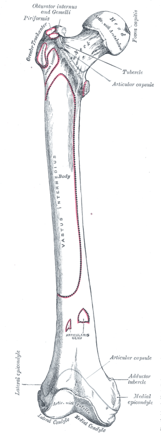
Femur, Right, Obturator internus, Gemelli, Piriformis, Greater Trochanter, Fovea capitis, Tubercle, Vastus Lateralis, Vastus Intermedius, Articularis Genu, Adductor tubercle, Medial epicondyle, Medial Condyle, Lateral Condyle, Patella,
Contributed by Gray's Anatomy Plates