Anatomy, Skeletal Muscle
- Article Author:
- Heeransh Dave
- Article Author:
- Micah Shook
- Article Editor:
- Matthew Varacallo
- Updated:
- 9/22/2020 1:48:19 PM
- For CME on this topic:
- Anatomy, Skeletal Muscle CME
- PubMed Link:
- Anatomy, Skeletal Muscle
Introduction
The musculoskeletal system comprises one of the major tissue/organ systems in the body. The three main types of muscle tissue are skeletal, cardiac and smooth muscle groups.[1][2][3] Skeletal muscle attaches to the bone by tendons, and together they produce all the movements of the body. The skeletal muscle fibers are crossed with a regular pattern of fine red and white lines, giving the muscle a distinctive striated appearance. Hence they are also known as striated muscle.[4][5][6][7][8]
Structure and Function
The skeletal muscle is one of the three significant muscle tissues in the human body. Each skeletal muscle consists of thousands of muscle fibers wrapped together by connective tissue sheaths. The individual bundles of muscle fibers in a skeletal muscle are known as fasciculi. The outermost connective tissue sheath surrounding the entire muscle is known as epimysium. The connective tissue sheath covering each fasciculus is known as perimysium, and the innermost sheath surrounding individual muscle fiber is known as endomysium.[9] Each muscle fiber is comprised of a number of myofibrils containing a number of myofilaments. When bundled together, all the myofibrils get arranged in a unique striated pattern forming sarcomeres which are the fundamental contractile unit of a skeletal muscle. The two most significant myofilaments are actin and myosin filaments which are arranged distinctively to form various bands on the skeletal muscle. The stem cells which differentiate into mature muscle fibers are known as satellite cells which can be found between the basement membrane and the sarcolemma (the cell membrane surrounding the striated muscle fiber cell).[10] When stimulated by growth factors, they differentiate and multiply to form new muscle fiber cells.[11]
The primary functions of the skeletal muscle take place via its intrinsic excitation-contraction coupling process. As the muscle is attached to the bone tendons, the contraction of the muscle leads to movement of that bone that allows for the performance of specific movements. The skeletal muscle also provides structural support and helps in maintaining the posture of the body. The skeletal muscle also acts as a storage source for amino acids that can be used by different organs of the body for synthesizing the organ-specific proteins.[12] The skeletal muscle also plays a central role in maintaining thermostasis and acts as an energy source during starvation.[9]
Embryology
Distinct transcriptional mechanisms and specific gene regulatory activity control the differentiation of muscle fibers.[13]During embryogenesis, it is the para-axial mesoderm that undergoes stepwise differentiation to generate the muscle tissue. The para-axial mesoderm on either side of the neural tube starts to differentiate and undergoes segmentation to form the somites. The somites get stimulated by the myogenic regulatory factors to differentiate into a dermomyotome and sclerotome. These regulatory factors include the Wnt, Shh and BMP4 proteins. The neural tube and the surface ectoderm are the primary sources of Wnt proteins, the Shh proteins(Sonic Hedge Hog) source from Notochord, and the lateral mesoderm plate produces the BMP4 protein.[14] The lateral aspect of the dermomyotome undergoes epithelial to mesenchymal transition as it proceeds to migrate on the ventral side to form a unique myotome below the dermatome.
The myotome then differentiates to form the skeletal muscles in the body after receiving stimulation from the Sonic Hedgehog (Shh) signaling molecule from the notochord that results in the Myf5 expression and subsequent differentiation.[15] The dorsomedial aspect of the myotome differentiates into epaxial myotome giving rise to back muscles. The ventrolateral aspect differentiates into hypaxial myotome that gives rise to muscles of the body wall.
Several signaling molecules like the Wnt and the BMP along with some transcription factors like Sine Oculis homeobox are responsible for this differentiation. The development of skeletal muscles in the limb and the trunk depends on the expression of MyoD and Myf5 and their effects on the different myoblasts.[16] These embryonic myoblasts undergo further differentiation to form the primary muscle fibers and eventually secondary myofibers by the union of myoblasts in the fetus. After birth, the satellite cells act as stem cells and are responsible for the further growth and development of skeletal muscles.
Blood Supply and Lymphatics
The main artery or the primary artery supplying blood to the skeletal muscle courses parallel to the longitudinal axis of the muscle fiber.[17] The primary artery gives off tributaries which are known as feed arteries, that are perpendicular to the primary artery and proceed towards the external connective tissue sheath of the muscle fiber called perimysium.[18] The feed artery branches into primary arterioles which after two more orders of branching gives rise to transverse arterioles, which in turn give rise to terminal arterioles.[19] The terminal arterioles are the final vascular branches, and they perfuse the capillaries that are present within the endomysium and travel parallel to the longitudinal axis of the muscle fiber. The terminal arteriole along with the capillaries that it supplies is known as a microvascular unit, and it is the smallest unit in the entire skeletal muscle where the blood flow can be regulated.
Lymph capillaries originate in skeletal muscle in the microvascular unit within the endomysium near the main capillary bed and drain the tissue fluid. These capillaries merge with each other to form the lymphatic vessels as they drain out the tissue fluid. These lymphatic vessels go through the perimysium and join with the larger lymphatic vessels. Unlike the blood vessels, the wall of the lymph vessels within the muscle does not have contractile property due to lack of smooth muscles(in the wall), so they depend on the muscle movement and arteriolar pulsations to drain the lymph out.
Nerves
The neuronal innervation of a skeletal muscle typically comprises of sensory nerve fibers, motor nerve fibers, and the neuromuscular junction. The nerve fibers are composed of myelinated as well as non-myelinated nerve fibers. The cell bodies of the neurons give rise to large axons which are generally unbranched and travel to the target muscles for innervation. Near the target muscle, the axons divide into multiple smaller branches to innervate multiple muscle fibers. The motor nerve terminal has abundant mitochondria, endoplasmic reticulum and numerous membrane-bound synaptic vesicles containing neurotransmitter- acetylcholine.[20] Once the action potential travels to the neuromuscular junction, there occurs a series of processes culminating into the fusion of the membrane of the synaptic vesicles with the presynaptic membrane and subsequent release of the neurotransmitter into the synaptic cleft.[21] [22]
The postsynaptic membrane of the muscle fibers has a massive concentration of neurotransmitters (AchR) receptors. These receptors are transmembrane ligand-gated ion channels.[23] Once the neurotransmitter activates these ion channels, there is a rapid depolarization of the motor end plate, which initiates an action potential in the muscle fiber resulting into muscle contraction.[21]
Muscles
Each muscle comprises multiple tissues including blood vessels, lymphatics, contractile muscle fibers, and the connective tissue sheaths. The outermost sheath of connective tissue covering each muscle is called as epimysium. Each muscle is made up of groups of muscle fibers called fascicles which are surrounded by a connective tissue layer called perimysium. Within each fascicle, there are multiple units of individual muscle fibers surrounded by endomysium, a connective tissue sheath. The two most essential myofilaments that make up the contractile elements of the muscle fiber are the actin and myosin. They are arranged distinctively in a striated pattern to form the dark A band, the light I band as well as the fundamental unit of contraction also referred to as a sarcomere. The sarcomere consists of a central M line and attached to it on either side are the thick myofilaments of myosin. This forms the dark A band. The sarcomere is bordered by the Z line that serves as the site of origin of the thin myofilaments of actin that project towards each other as they partially overlap the myosin filaments.[9]The regulatory proteins namely troponin C, I, T as well as tropomyosin play a key role in the myofilaments sliding mechanism leading to contraction. Titin and nebulin are the other major proteins that contribute to the mechanical properties of the muscle.[24] There is a unique T-tubule system in place for conduction of neuronal action potential to the interior of the muscle cell via invaginations of the sarcolemma to enhance coordination and uniform muscle contraction.[25]
Clinical Significance
Skeletal muscles enable humans to move and perform daily activities. They play an essential role in respiratory mechanics and help in maintaining posture and balance. They also protect the vital organs in the body.
Various medical conditions occur as a result of abnormalities in the function of skeletal muscles. Some of these diseases include myopathies, paralysis, myasthenia gravis, urinary and or bowel incontinence, ataxia, weakness, tremors, among others. Disorders of the nerves can cause neuropathy and cause disturbances in the functionality of the skeletal muscles as well. In addition, skeletal muscle/tendon ruptures can occur acutely in high-level athletes or recreational sports participants and generate significant disability in all patients regardless of activity status.[26]
Muscle Cramps
Muscle cramps result in continuous, involuntary, painful, and localized contraction of an entire muscle group, individual single muscle, or select muscle fibers.[3] Generally, the cramp can last from minutes to a few seconds for idiopathic or known causes with healthy subjects or in the presence of diseases. Palpating the muscle area of the cramp will present a knot.
Exercise-associated muscle cramps are the most frequent condition requiring medical/therapeutic intervention during sports.[27] The specific etiology is not well understood, and possible causes depend on the physiological or pathological situation in which the cramps appear. It is important to note that a painful contraction that is limited to a specific area does not mean that the cause of the onset of the cramp is necessarily local.
In specific clinical scenarios, the underlying etiology may relate to persistent, spastic muscle contractions that significantly can impact human function. A typical example of this condition manifests in the sternocleidomastoid muscle. Clinically, this is recognized in congenital torticollis or spasmodic torticollis.[28]
Other relevant conditions in this realm include, but are not limited to the following:
- Exercise-induced and heat-related muscle cramping
- Piriformis syndrome[6][29]
- Thoracic outlet syndrome (scalene muscle hypertrophy/spasticity)[5]
Palsy/Compression Neuropathy
At the opposite end of the spectrum, various muscle palsies exist secondary to the long-term, downstream effects of various nerve conditions and neuropathies that can result in frankly flaccid conditions (which may be permanent or temporary). These syndromes and conditions include, but are not limited to the following:
(Click Image to Enlarge)
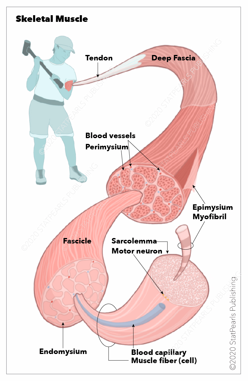
Skeletal muscles, Sarcolemma, Myofibril, Motor neuron, Blood capillary, Endomysium, Muscle fiber (cell), Fascicle, Perimysium, Blood vessels, Epimysium, Tendon, Deep Fascia
Illustration by Emma Gregory