Thromboelastography
- Article Author:
- Maxim Shaydakov
- Article Author:
- David Sigmon
- Article Editor:
- John Blebea
- Updated:
- 4/24/2020 10:03:29 PM
- For CME on this topic:
- Thromboelastography CME
- PubMed Link:
- Thromboelastography
Introduction
Maintaining blood in a liquid state is critical for homeostasis. It allows blood to supply an adequate delivery of oxygen and nutrients to tissues while also eliminating carbon dioxide and other waste products. On the other hand, the ability of blood to convert from a liquid to a solid state, in other words, to coagulate, underlies the mechanism that protects the body from life-threatening exsanguination. This process of thrombosis is normally a localized event at the site of vascular injury while the rest of the circulating blood remains in a liquid state. Thrombosis is a dynamic process that includes associated thrombolysis to maintain or restore blood flow through vessels once an injury has been sealed. These unique properties of blood are largely determined by a complex and active balance between pro-coagulation factors, anticoagulants, and fibrinolysis. Two major pathologic conditions are commonly associated with a disequilibrium of this intricate system: bleeding and vessel thrombosis.
Major bleeding is a serious medical complication that may be caused by external trauma, surgery, invasive procedures, or an underlying medical condition such as aneurysm rupture or peptic ulcer disease. According to the World Health Organization (WHO), injuries are responsible for 5.8 million deaths per year worldwide, with the associated bleeding responsible for about 30% to 40% of these deaths.[1] A number of congenital disorders associated with a coagulation factor deficiency, such as Von Willebrand disease, hemophilia A or B, may cause significant bleeding even with minor injuries. In addition, prescribed anticoagulants and antiplatelet agents may create a coagulopathic state that may lead to excessive bleeding either associated with trauma or medical procedures. Finally, major acute blood loss can lead to coagulopathy due to a loss of coagulation factors. Predictably, trauma-related coagulopathy has been associated with significantly higher mortality.[2] Patients with ongoing or expected major bleeding would benefit from an accurate assessment of the functional state of the hemostatic system to provide optimal care, providing cost-effective replacement of only the needed blood components.
Venous thromboembolism (VTE) is another common and serious condition that is associated with abnormal blood coagulation. In these cases, systemic hypercoagulability shifts the body’s homeostatic mechanisms toward a pro-thrombotic state. In particular patients, however, a definitive cause for the VTE may be unclear. Routine coagulation testing has not been shown to predict such events, and in many cases, even a detailed hypercoagulability investigation fails to identify an underlying disorder. A large number of people take anticoagulants and antiplatelet agents on a regular basis[3][4] which impacts the accuracy of the results of many laboratory coagulation studies. An accurate and cost-efficient method of monitoring antithrombotic activity would be helpful to maintain an acceptable risk/benefit ratio in such patients. Inadequate anticoagulation or antiplatelet therapy can lead to devastating thromboembolic conditions.
Several commonly used blood tests assess blood coagulation. These tests include prothrombin time (PT), international normalized ratio (INR), activated partial thromboplastin time (aPTT), platelet count, fibrinogen concentration, D-dimer level, activated clotting time, and whole blood bleeding time (BT). These tests are usually used for the clinical diagnosis of coagulopathy and a possible prothrombotic state, to monitor anticoagulation therapy, and to assist in the treatment of bleeding episodes. More specific factor analyses, such as Factor V, proteins C and S, anti-thrombin III, anticardiolipin antibodies and prothrombin gene mutation are useful but not as readily available in emergency clinical situations. Despite being very effective for specific clinical needs, such as anticoagulation monitoring, the first group of usual diagnostic tests has limitations. Their main disadvantage in circumstances of acute major bleeding is long turnaround time. Furthermore, they do not provide a complete picture of hemostasis due to their inability to assess some coagulation factors (such as Factor XIII), platelet function and the activity of the fibrinolytic system. Platelet concentration, easily measured as part of a complete blood count, does not necessarily reflect their function, especially in the presence of elements known to affect platelet reactivity, such as non-steroidal anti-inflammatory drugs, antiplatelet agents, uremia, malignancy, or alcohol intake. Bleeding time has a low sensitivity and high inconsistency in detecting platelet disorders.[5] Delayed or inadequate diagnosis of coagulopathy in a bleeding patient may lead to an excessive and improperly balanced transfusion of scarce blood components with increased morbidity, treatment costs, and mortality.[6]
Thromboelastography (TEG) is a promising diagnostic modality that offers several advantages compared to the other tests that have been mentioned above. TEG was developed and first described by Dr. Hellmut Hartert at the University of Heidelberg (Germany) in 1948.[7] The first reported clinical application of the test occurred during the Vietnam War in an attempt to guide transfusions of blood components in injured soldiers.[8] In 1980s, TEG was found to be beneficial in liver transplant patients[9], and in 1990s, was demonstrated to be useful in cardiac surgery.[10] Since then, TEG has evolved into a more commonly used test as more evidence for its clinical efficacy has been attained. A brief search in PubMed using keywords “thromboelastography” and “thromboelastometry” results in about 6000 publications. This article will describe the general principles of TEG, methodology, normal values, along with the current evidence and clinical applications, as well as limitations and future research directions.
Pathophysiology
TEG is a non-invasive test that quantitatively measures the ability of whole blood to form a clot. The principle of this in vitro test is to detect and quantify dynamic changes of the viscoelastic properties of a blood sample during clotting under low shear stress. The test is performed in a specially designed system called a thromboelastograph. The system consists of 2 chambers simultaneously examining a blood sample in duplicate to reduce the risk of sampling and measurement errors. Each chamber consists of a platform that holds a disposable cup where a blood sample is placed and a detection pin suspended in its center. The cup oscillates around the detection pin in a limited arc of plus or minus (+/-) 4 degrees 45' every 5 seconds. Induced pin movement is recorded and changes measured as a function of time. Initially, there is little movement of the pin since liquid blood possesses minimal viscosity and the oscillations of the cup are not transmitted to the pin. As the blood coagulates, it begins to adhere to both the cup and the pin and movement of the cup induces motion on the pin. These gradually increasing viscoelastic mechanical properties of the blood are reflective of the developing 3-dimensional fibrin mesh and platelet components of the clot. The greater the viscoelasticity of the clot, the higher the amplitude of the pin motion. As fibrinolysis starts, the fibrin-platelet structure begins to dissolve gradually, and the clot loses its contact with the detection pin which has less induced motion. The thromboelastogram (Figure 1) is a graphical image of the recorded amplitude of movement of the pin as a function of time. Analytical software measures and quantifies these changes. Therefore, TEG measures the functional ability of the blood to make a hemostatic plug. A newer version replaces the cup rotation method with a resonance technique wherein the blood sample is subjected to vibration, and the vertical movement of the blood meniscus is measured under LED illumination. The system uses pre-measured cartridges that do not require pipetting and allows simultaneous performance of four blood tests.
Specimen Requirements and Procedure
The blood sample is collected via venipuncture in a plastic vial with 3.2% buffered sodium citrate with a citrate-to-blood ratio of 1:9. The vial is inverted several times to mix the blood and citrate. Maintaining this citrate-blood ratio is crucial for test accuracy. Citrate binds calcium, an important cofactor of coagulation, preventing the blood from clotting before the beginning of the test. A clotted specimen, reflecting a vial overfilled with blood, cannot be used. For TEG testing, the collected non-clotted samples are considered stable and usable for up to 2 hours at room temperature. Non-citrated whole blood (native blood TEG or NATEM) can also be tested, but it must be used immediately. The test and reagents used are at room temperature. A volume of 340 uL of citrated blood is pipetted to the study cup, recalcified by the addition of 20 uL of 0.2M calcium chloride and then activated with a kaolin-cephalin reagent. Cephalins, or phosphatidylethanolamines, are a class of phospholipids commonly present in membranes of human cells. They are an important cofactor of the coagulation cascade which enables the assembly of tenase and prothrombinase complexes on the surface of platelets which are critical for thrombin generation. Kaolin is a mineral, primarily composed of hydrated aluminum silicate, which is a negatively charged molecule that can initiate the intrinsic coagulation pathway by activating Factor XII. Precise proportioning of the blood and kaolin-cephalin reagent is important for accurate and reproducible TEG results.[11] Non-activated TEG is also possible, but the lack of activators significantly prolongs clotting time and the testing process which is not desirable in a clinical emergency.
There have been several modifications of classic TEG assay that have been developed to improve its diagnostic value. Rapid TEG (r-TEG) utilizes tissue factor instead of the kaolin-cephalin reagent to activate blood coagulation. Because tissue factor triggers the extrinsic coagulation pathway which involves a smaller number of coagulation factors, the test can be performed faster than conventional TEG. Rapid TEG can be completed within 15 minutes and thus is helpful in managing massive transfusions in trauma patients.[12][13] The TEG platelet mapping assay was developed to predict the inhibitory effect of antiplatelet agents such as aspirin and clopidogrel. This is accomplished by evaluating platelet aggregation in the presence of adenosine diphosphate or arachidonic acid. TEG with added heparinase (hTEG) measures the effect of heparin reversal on blood coagulation.
Rotational thromboelastography, also known as rotational thromboelastometry (RoTEM) utilizes an oscillating pin which rotates +/- 4 degrees 45' every 6 seconds while maintaining the cup in a stable position. In this assay, some different activator reagents are utilized to investigate specific components of the coagulation pathway (Table 1).
Results, Reporting, Critical Findings
A normal thromboelastogram is schematically represented in Figure 1. Prompt qualitative analysis of the TEG tracing can be performed during the test. The quantitative analysis of TEG includes the measurement of the 5 parameters listed and described in Table 1. A coagulation index has also been suggested by the manufacturer to assess the overall coagulation status. The coagulation index (CI) for whole blood may be calculated as follows:
- CI = -0.2454R+ 0.0184K + 0.1655MA - 0.0241a - 5.0220
Normal values of the coagulation index lie within -3.0 and +3.0, which is three standard deviations from the mean of zero. A hypercoagulable state is defined as CI greater than +3.0 and coagulopathy as CI less than -3.0. Previous studies demonstrated a significantly elevated CI in postoperative period after general surgery and in cancer patients, suggesting a prothrombotic state.[14][15] However, this index is not widely used and its clinical usefulness not yet validated. Several other parameters may be calculated based on the thromboelastogram, such as projected maximal amplitude, time to maximal amplitude, the G parameter (shear elastic modulus strength, or clot strength), and a thrombodynamic potential index. While providing interesting information these variables are rarely used in clinical practice.
Normal TEG values are presented in the Table 2, although some patient-related factors may affect these values. Elderly patients demonstrate a tendency towards more pro-coagulable TEG results suggesting a need for correction of these reference values in elderly.[16] In some circumstances, such as in patients undergoing cardiac surgery and liver transplantation, specific reference values are less important because the principal application of TEG is to compare the patient’s own baseline to changes during the intraoperative and postoperative periods. In other clinical scenarios, such as trauma or postoperative bleeding, reference values are important for interpretation of the results as no baseline data is available. There is also some variability in testing results. A study on 118 healthy volunteers revealed at least one abnormal parameter in 19%, and a coagulopathy (defined as at least 2 abnormal parameters) in 9%, leading to a calculated specificity of 81%.[17] A larger prospective trial of more diverse group of subjects would be helpful to establish analyzer-specific and reagent-specific reference values in selected subgroups of patients.
Deviation of each of these TEG parameters from the reference values suggests specific disturbances of hemostasis and coagulation. Prolongation of the R time reflects a quantitative or qualitative deficiency of coagulation factors that may be corrected by fresh frozen plasma (FFP) transfusion, prothrombin complex, or anticoagulant reversal. Prolongation of the K time, or a decrease of the alfa angle, suggests a deficiency of fibrinogen and may be corrected by cryoprecipitate or lyophilized fibrinogen concentrate. Low MA indicates a quantitative or functional deficiency of platelets and could be corrected by platelet concentrate transfusion or desmopressin. Finally, an increased LY value implies an activated fibrinolysis that may be treated by fibrinolysis inhibitors (aminocaproic or tranexamic acid). The opposite changes in TEG parameters suggest a prothrombotic state. This interpretive approach represents a convenient but rather simplified view on disturbances of blood coagulation. It is important to remember that due to the complex nature of hemostasis these TEG parameters are interrelated.
A different nomenclature is used for RoTEM assays to define the same TEG parameters: clotting time (CT) instead of R, clot formation time (CFT) instead of K, maximum clot firmness (MCF) instead of MA, and CL instead of LY. Reference values for RoTEM have been established in a multicenter study on 500 healthy volunteers.[18] Depending on the specific TEG analyzer (TEG versus RoTEM) and reagents being used, differing results may be obtained from the same blood sample, potentially affecting clinical decision-making.[19],[20] Some of these differences are not well understood. For example, NATEM may be more sensitive to hyperfibrinolysis than INTEM and EXTEM.[21] A systematic review of four clinical trials comparing TEG and RoTEM found clinically significant differences between the 2 tests with a lack of comparability of the results.[22] A recent systematic review did not discover a sufficient number of well-designed studies to be able to compare the results of TEG and RoTEM in healthy subjects.[23] Hence, the results of different modifications of TEG and RoTEM cannot be considered interchangeable until head-to-head prospective comparative studies are performed.
Clinical Significance
The main advantage of TEG testing is its potential to deliver immediate goal-oriented and individualized care to a bleeding patient:
- Global assessment of blood coagulability, including coagulation cascade, platelet function, and fibrinolysis
- Rapid real-time bedside test with a simple methodology (point-of-care testing)
- Diagnosis of coagulopathic bleeding
- Guide transfusion therapy and decrease the use of blood products
- Detect dynamic changes in blood coagulation during resuscitation
- Predict the clinical efficacy of therapeutic agents affecting blood coagulability
TEG has convincingly demonstrated its usefulness to help improve outcomes in cardiac surgery. A meta-analysis of 17 randomized controlled trials (RCTs) demonstrated that TEG decreases blood product transfusions and surgical re-exploration due to postoperative bleeding in cardiac surgery patients.[24] These effects were associated with a lower incidence of acute kidney injury and thromboembolic events. Another systematic review of 17 RCTs involving 1493 patients, mainly elective on-pump cardiac surgery, revealed that TEG/RoTEM decreases transfusion of blood components and reduces the overall mortality.[25] The quality of the included studies, however, was considered to be low.[25] A recent RCT found that intraoperative correction of coagulopathy guided by EXTEM and FIBTEM can reduce postoperative bleeding, blood transfusions, and duration of critical care in pediatric cardiac surgery patients.[26] TEG is also a more cost-effective method compared to standard coagulation tests in the diagnosis of coagulopathy in cardiac surgery.[27]
There is conflicting evidence for TEG usefulness in trauma patients. A recent Cochrane database systematic review found insufficient data to compare the accuracy of TEG and RoTEM versus PT/INR in the diagnosis of trauma-induced coagulopathy.[28] The review concluded that these tests are still in the phase of clinical research. However, it is questionable whether PT/INR can be considered a good reference standard to diagnose coagulopathy. In major trauma, r-TEG has been found to be better in predicting the need for transfusion of FFP, RBCs, and platelets compared to conventional coagulation tests of PT, aPTT, INR, platelet count, and fibrinogen.[29] Based on another large systematic review, although with evidence limited to cohort studies with a moderate to high risk of bias, TEG/RoTEM can diagnose coagulopathy and may predict blood components transfusion and mortality in trauma patients.[30] Another review of 13 cohort studies involving only RoTEM in 2835 adult trauma patients came to the same conclusions.[31] However, there was no improvement in patient morbidity or mortality.[30] Another Cochrane database systematic review of 9 RCTs with a total of 776 participants, mainly cardiac surgery patients, found a decreased amount of bleeding when TEG or RoTEM were utilized but also without a decrease in morbidity or mortality.[22] This inability of TEG/RoTEM testing to significantly reduce mortality may become a barrier to widespread clinical use. However, it is important to realize that overall mortality in hospitalized patients with bleeding is relatively low and thus would require large clinical trials to detect a statistically significant impact of TEG on mortality. Furthermore, these are complex patients and the overall treatment strategy, rather than diagnostic testing, will have a greater role in affecting overall morbidity and mortality.
There is expanding evidence of using different TEG and RoTEM assays in other additional clinical scenarios. A small RCT has demonstrated the ability of TEG to better guide anticoagulation during ECMO compared to aPTT, reducing the dose of heparin.[32] TEG platelet mapping can detect platelet inhibition by clopidogrel and aspirin in surgical patients.[33] A novel TEG-based scoring system has been suggested to diagnose disseminated intravascular coagulation.[34] TEG may detect possible coagulopathy in patients with intracranial bleeding and hematoma enlargement.[35] TEG may also have an application in liver disease patients. It is known that conventional coagulation tests are commonly abnormal in liver disease. At the same time, TEG/RoTEM results are normal in many patients despite an abnormal INR or platelet count due to adjustments in the system of hemostasis, or rebalanced hemostasis.[36] Thus, TEG/RoTEM may provide a better insight into the risk of bleeding in patients with liver disease than conventional coagulation tests.[37] EXTEM has been found helpful to detect intraoperative coagulopathy in liver transplant patients.[38] There are other clinical situations when TEG has demonstrated potential benefit, but listing all of them is beyond the scope of this review.
Clinical Guidelines
Thromboelastography is recommended by NICE guidelines to help detect, manage and monitor hemostasis in cardiac surgery patients (NICE guidelines, 2014). Other clinical guidelines do not currently strongly recommend TEG for use in other settings due to the lack of high-quality evidence. Recently updated guidelines of the European Society of Anesthesiology recommended viscoelastic hemostatic assays (TEG/RoTEM) to guide the management of perioperative bleeding and for managing severe peripartum hemorrhage albeit with the low level of evidence.[39]
Limitations
An ideal test on blood coagulation does not yet exist. TEG measures blood coagulation in vitro, with or without an additional activator. An important component of the coagulation cascade, tissue factor, cannot be quantified in vitro. Moreover, blood coagulation potential is only one component in such complex processes as clinical thrombosis and bleeding. Blood coagulation also depends on the size of the injured vessel, blood flow characteristics, and local vessel wall biology that determines the quantity and functional activity of the membrane-bound pro- and anticoagulation factors. In other words, there are significant aspects of coagulation which are not components of the blood. An abnormal TEG in a patient without clinically relevant bleeding does not require transfusion of blood components. A single test or patient-related factor rarely guide the decision to transfuse blood components or initiate/correct antithrombotic therapy.
TEG has a sensitivity and specificity that may vary significantly in different populations. Patients taking anticoagulants and antiplatelet agents are a major concern the trauma setting. Warfarin is a commonly prescribed medication [40] that has been associated with increased mortality in trauma patients [41]. In about half of patients on warfarin therapy, R-time may be normal in both TEG and rapid TEG tests, with a poor correlation between TEG and INR.[11] This is a good example of how TEG may miss a clinically significant coagulopathic state. Hence, INR is still the gold standard of monitoring warfarin therapy. Several important blood tests also cannot be currently replaced by TEG, such as P2Y12 platelet function assay to guide clopidogrel therapy, D-Dimer to exclude VTE in low-risk outpatients, and advanced thrombophilia diagnostic tests.
Future Directions: Guiding Anticoagulation and Antiplatelet Therapy
Anticoagulation therapy is a field where TEG may become more applicable pending future clinical studies. Up to one-third of patients on warfarin therapy may have subtherapeutic anticoagulation at some point of treatment.[42] One of the advantages of direct oral anticoagulants (DOAKs) is no requirement to monitor anticoagulation therapy. However, they may be problematic in certain groups of patients, such as those with renal failure, liver failure, pregnancy, extremes of body weight, high bleeding risk, thrombosis progression or recurrence on anticoagulation.[43] High plasma concentration of DOAKs has been associated with higher bleeding risk.[44],[45] It these situations, TEG may provide the ability to adjust the level of anticoagulation during the same office visit. Since TEG implies activation of an intrinsic coagulation pathway with kaolin, it is sensitive to heparin and low molecular weight heparin therapy (R time). An interesting case of an emergent heparin reversal under TEG control in a patient with intracranial bleeding has been reported.[46]
Prediction of platelet inhibition by antiplatelet agents (such as aspirin, clopidogrel, abciximab, eptifibatide, or tirofiban) is another promising avenue for TEG application. Most antiplatelet agents are used with a standard dose despite several known issues associated with this approach. For example, up to 25% of patients with STEMI may be resistant to clopidogrel which increases the risk of recurrent cardiovascular events.[47] Aspirin resistance has been associated with an increased incidence of myocardial infarction, stroke or death in patients with cardiovascular disease.[48] Hence, there is significant variability in individual response to antiplatelet therapy. Available evidence does not support the use of usual laboratory testing to guide the dose of aspirin or clopidogrel.[49] Future studies may determine if TEG can measure the effect of antiplatelet therapy, detect hyporesponsiveness, and predict the risk of bleeding or thromboembolic complications.
A novel concept of individualized health care applies to both anticoagulation and antiplatelet therapy monitoring. Using a standard dose of the same medications to treat patients with different medical conditions and comorbidities may not be an ideal approach. The potential of TEG to improve the quality of antithrombotic therapy is a promising avenue for experimental and clinical research.
Future Directions: Prevention of Venous Thromboembolism
Another potential application of TEG is to improve diagnosis, prevention, and treatment of patients with venous thromboembolism. The most appropriate evidence-based practice to prevent VTE is to stratify patients based on the VTE risk using one of the risk prediction models elaborated for surgical and medical patients. None of these models includes conventional blood coagulation tests since they do not predict VTE. A number of initial reports suggest that TEG may be a useful tool to help with risk stratification. TEG may have a VTE predictive value in critically ill patients[50][51], gynecological oncology patients[52], and prostate cancer [53]. A large prospective cohort of adult trauma patients revealed a 2-fold higher risk of VTE in patients with hypercoagulable TEG parameters on arrival to the trauma bay.[54] However, other reports did not find TEG of value in predicting VTE in selected patients, such as orthopedic surgery.[55]
Despite an appropriate prophylaxis, VTE is still a frequent concern in hospitalized patients. In one study, R time of TEG was significantly shorter in critically ill patients on LMWH prophylaxis who develop DVT compared to those patients who do not.[56] Thus, TEG may be helpful to predict VTE that occurs despite standard pharmacological prophylaxis.
Quality control and Lab Safety
(Click Image to Enlarge)
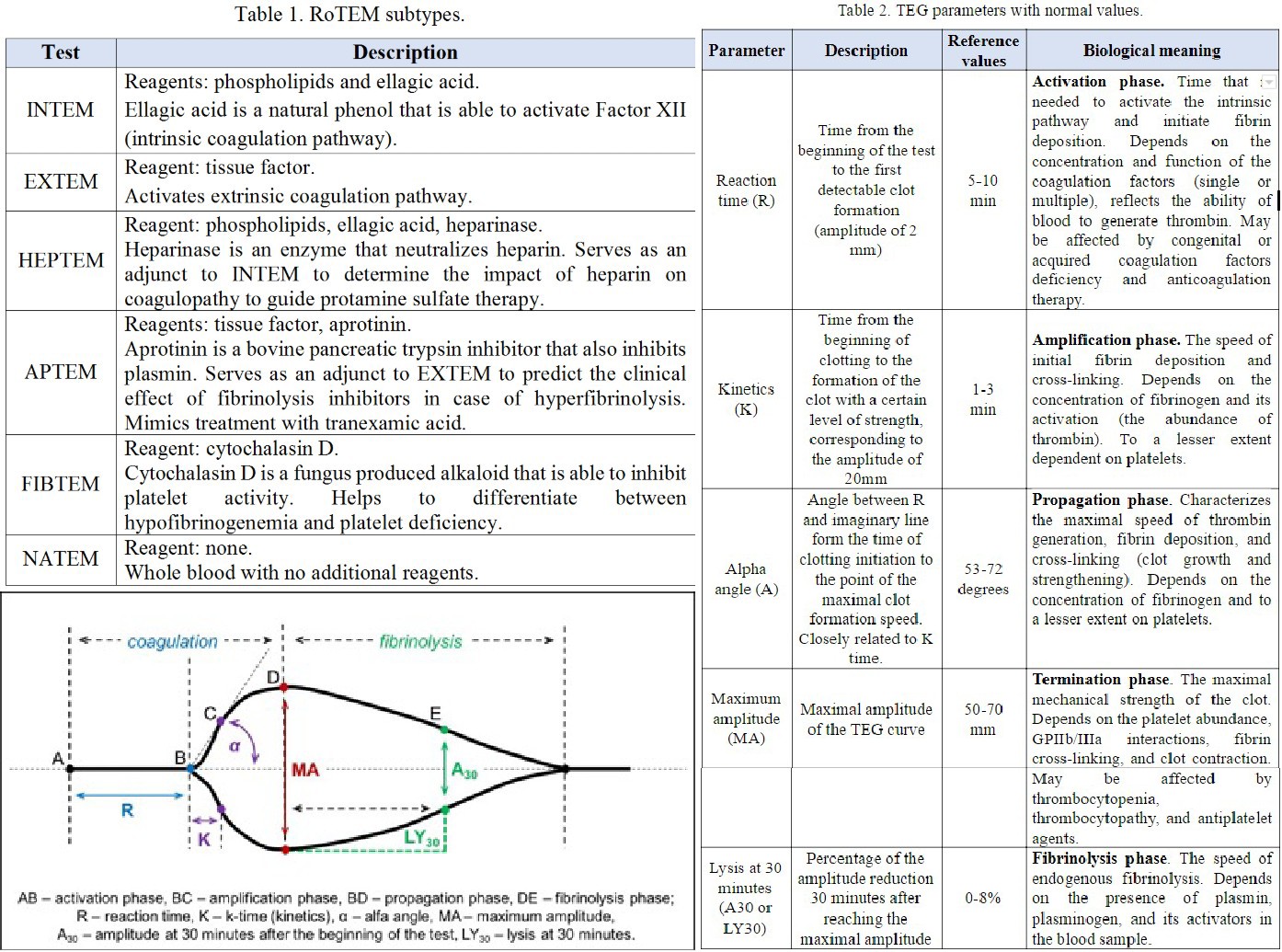
Theombelastography Tables
Contributed by Maxim Shaydakov