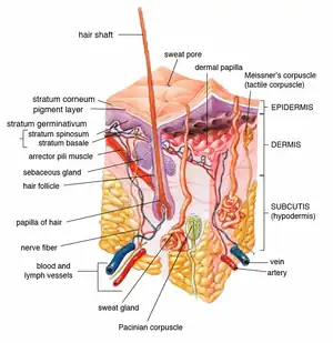
Arrector pili muscle
| Details | |
|---|---|
| Nerve | Sympathetic postganglionic nerve fibers |
| Identifiers | |
| TA98 | A16.0.00.024 |
| TA2 | 7051 |
| TH | H3.12.00.3.01041 |
| FMA | 67821 |
| Anatomical terms of muscle | |
The arrector pili muscles, also known as hair erector muscles,[1] are small muscles attached to hair follicles in mammals. Contraction of these muscles causes the hairs to stand on end,[2] known colloquially as goose bumps (piloerection).[3]
Structure
Each arrector pili is composed of a bundle of smooth muscle fibres which attach to several follicles (a follicular unit).[4] Each is innervated by the sympathetic division of the autonomic nervous system.[4] The muscle attaches to the follicular stem cell niche in the follicular bulge,[3][4][5] splitting at their deep end to encircle the follicle.[6]
Function
The contraction of the muscle is involuntary. Stresses such as cold, fear etc. may stimulate the sympathetic nervous system, and thus cause muscle contraction.[4]
Thermal insulation
Contraction of arrector pili muscles have a principal function in the majority of mammals of providing thermal insulation.[4] Air becomes trapped between the erect hairs, helping the animal retain heat.
Self defence
Erection of the porcupine's long, thick hairs causes the animal to become more intimidating, scaring predators.
Sebum excretion
Pressure exerted by the muscle may cause sebum to be forced along the hair follicle towards the surface, protecting the hair.[7]
Hair follicle stability
Arrector pili muscles also stabilise the base of the hair follicle.[5][6]
Clinical significance
Skin conditions such as leprosy can damage arrector pili muscles, preventing their contraction.[8]
History
The term "arrector pili" comes from Latin. It translates to "hair erector".[1]
Additional images
 Insertion of sebaceous glands into hair shaft
 Cross-section of all skin layers
Notes
- 1 2 "Anatomy of the Skin | SEER Training". training.seer.cancer.gov. Retrieved 2021-01-21.
- ↑ David H. Cormack (1 June 2001). Essential histology. Lippincott Williams & Wilkins. pp. 1–. ISBN 978-0-7817-1668-0. Retrieved 15 May 2011.
- 1 2 Fujiwara, Hironobu; Ferreira, Manuela; Donati, Giacomo; Marciano, Denise K.; Linton, James M.; Sato, Yuya; Hartner, Andrea; Sekiguchi, Kiyotoshi; Reichardt, Louis F.; Watt, Fiona M. (2011-02-18). "The Basement Membrane of Hair Follicle Stem Cells Is a Muscle Cell Niche". Cell. 144 (4): 577–589. doi:10.1016/j.cell.2011.01.014. ISSN 0092-8674. PMC 3056115. PMID 21335239.
- 1 2 3 4 5 Pascalau, Raluca; Kuruvilla, Rejji (August 2020). "A Hairy End to a Chilling Event". Cell. 182 (3): 539–541. doi:10.1016/j.cell.2020.07.004. ISSN 0092-8674. PMID 32763185. S2CID 221012408.
- 1 2 Torkamani, Niloufar; Rufaut, Nicholas; Jones, Leslie; Sinclair, Rodney (2017-01-01). "The arrector pili muscle, the bridge between the follicular stem cell niche and the interfollicular epidermis". Anatomical Science International. 92 (1): 151–158. doi:10.1007/s12565-016-0359-5. ISSN 1447-073X. PMID 27473595. S2CID 26307123.
- 1 2 Poblet, Enrique; Jiménez, Francisco; Ortega, Francisco (August 2004). "The contribution of the arrector pili muscle and sebaceous glands to the follicular unit structure". Journal of the American Academy of Dermatology. 51 (2): 217–222. doi:10.1016/j.jaad.2004.01.054. ISSN 0190-9622. PMID 15280840.
- ↑ Journal of the American Academy of Dermatology Volume 51, Issue 2, August 2004, Pages 217-222 The contribution of the arrector pili muscle and sebaceous glands to the follicular unit structure☆ Enrique Poblet, Francisco Ortega. https://doi.org/10.1016/j.jaad.2004.01.054
- ↑ Budhiraja, Virendra; Rastogi, Rakhi; Khare, Satyam; Khare, Anjali; Krishna, Arvind (2010-09-01). "Histopathological changes in the arrector pili muscle of normal appearing skin in leprosy patients". International Journal of Infectious Diseases. 14: e70–e72. doi:10.1016/j.ijid.2009.11.018. ISSN 1201-9712. PMID 20207571.
References
- Myers, P., R. Espinosa, C. S. Parr, T. Jones, G. S. Hammond, and T. A. Dewey. 2006. The Animal Diversity Web; https://web.archive.org/web/20110903154915/http://animaldiversity.ummz.umich.edu/site/topics/mammal_anatomy/hair.html
- Burkitt, Young; et al. (1993). Wheater's Functional Histology: a text and colour atlas. Heath. p. 162.