Lacrimal nerve
| Lacrimal nerve | |
|---|---|
 Nerves of the orbit, the lacrimal nerve is visible, labelled over the eye. | |
| Details | |
| From | ophthalmic nerve |
| Innervates | lacrimal gland, conjunctiva, skin of lateral forehead, scalp and upper eyelid |
| Identifiers | |
| Latin | nervus lacrimalis |
| TA98 | A14.2.01.018 |
| TA2 | 6198 |
| FMA | 52628 |
| Anatomical terms of neuroanatomy | |
The lacrimal nerve is the smallest branch of the ophthalmic nerve (V1), itself a branch of the trigeminal nerve (CN V). The other branches of the ophthalmic nerve are the frontal nerve and nasociliary nerve.[1]
Structure
The lacrimal nerve branches from the ophthalmic nerve immediately before traveling through the superior orbital fissure to enter the orbit. It travels through it lateral to the frontal nerve outside the annulus of Zinn. After entering, it travels anteriorly along the lateral wall with the lacrimal artery, above the upper margin of the lateral rectus. It receives a communicating branch from the zygomatic nerve which carries the postganglionic parasympathetic axons from the pterygopalatine ganglion. It travels through the lacrimal gland giving sensory and parasympathetic branches to it and then continues anteriorly as a few small sensory branches.
Function
The lacrimal nerve provides sensory innervation to the lacrimal gland, conjunctiva of the lateral upper eyelid and superior fornix, the skin of the lateral forehead, scalp and lateral upper eyelid.
It also provides parasympathetic innervation to the lacrimal gland from the communicating branch it receives from the zygomatic nerve.
Additional images
 Superior view of the nerves of the orbit. The lacrimal nerve is seen branching from the ophthalmic nerve.
 Sensory innervation to the skin of the head and neck. The cutaneous distribution of the lacrimal nerve can be seen above the eye in the green area.
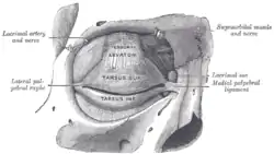
 Anterior view of the orbit and tarsal plates. The lacrimal nerve can be seen exiting the orbit superolaterally after it supplies the lacrimal gland.
 Superior view of a dissection of the left orbit. The lacrimal nerve is visible innervating the lacrimal gland.