Achilles tendinitis
| Achilles tendinitis | |
|---|---|
| Other names: Achilles tendinopathy, Achilles tendonitis, Achilles tenosynovitis | |
 | |
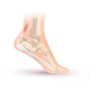
| Drawing of Achilles tendonitis with the affected part highlighted in red | |
| Specialty | Rheumatology |
| Symptoms | Pain, swelling around the affected tendon[1] |
| Usual onset | Gradual[1] |
| Duration | Months[2] |
| Types | Noninsertional, insertional[2] |
| Causes | Overuse[2] |
| Risk factors | Trauma, lifestyle that includes little exercise, high-heel shoes, rheumatoid arthritis, medications of the fluoroquinolone or steroid class[1] |
| Diagnostic method | Based on symptoms and examination[3] |
| Differential diagnosis | Achilles tendon rupture[3] |
| Treatment | Rest, ice, non-steroidal antiinflammatory agents (NSAIDs), physical therapy[1][2] |
| Frequency | Common[2] |
Achilles tendinitis, also known as achilles tendinopathy, occurs when the Achilles tendon, found at the back of the ankle, becomes inflamed.[2] The most common symptoms are pain and swelling around the affected tendon.[1] The pain is typically worse at the start of exercise and decreases thereafter.[3] Stiffness of the ankle may also be present.[2] Onset is generally gradual.[1]
It commonly occurs as a result of overuse such as running.[2][3] Other risk factors include trauma, a lifestyle that includes little exercise, high-heel shoes, rheumatoid arthritis, and medications of the fluoroquinolone or steroid class.[1] Diagnosis is generally based on symptoms and examination.[3]
While stretching and exercises to strengthen the calf are often recommended for prevention, evidence to support these measures is poor.[4][5] Treatment typically involves rest, ice, non-steroidal antiinflammatory agents (NSAIDs), and physical therapy.[1][2] A heel lift or orthotics and ultrasound treatment may also be helpful.[2][3][6] Local injections are generally not recommended due to risk of tendon rupture.[6] In those whose symptoms last more than six months despite other treatments, surgery may be considered.[2] Achilles tendinitis is relatively common.[2]
Signs and symptoms
Symptoms can vary from an ache or pain and swelling to the local area of the ankles, or a burning that surrounds the whole joint. With this condition, the pain is usually worse during and after activity, and the tendon and joint area can become stiffer the following day as swelling impinges on the movement of the tendon. Some thickening of the tendon can also occur.[2]
Cause


Achilles tendinitis is a common injury, particularly in sports that involve lunging and jumping.[7] It is also a known side effect of fluoroquinolone antibiotics such as ciprofloxacin, as are other types of tendinitis[8]
Achilles tendinitis is thought to have physiological, mechanical, or extrinsic (i.e. footwear or training) causes. Physiologically, the Achilles tendon is subject to poor blood supply through the synovial sheaths that surround it. This lack of blood supply can lead to the degradation of collagen fibers and inflammation.[9] Tightness in the calf muscles has also been known to be involved in the onset of Achilles tendinitis.[10]
During the loading phase of the running and walking cycle, the ankle and foot naturally pronate and supinate by approximately 5 degrees.[11] Excessive pronation of the foot in the subtalar joint is a type of mechanical mechanism that can lead to tendinitis.[10]
An overuse injury refers to repeated stress and strain, which is likely the case in endurance runners. Overuse can simply mean an increase in running, jumping or plyometric exercise intensity too soon.[12]
Risk factors
Risk factors include participating in a sport or activity that involves running, jumping, bounding, and change of speed; although Achilles tendinitis is mostly likely to occur in runners, it also is more likely in participants in basketball, volleyball, dancing, gymnastics and other athletic activities.[13] Other risk factors include gender, age, improper stretching, and overuse.[14] Another risk factor is any congenital condition in which an individual's legs rotate abnormally, which in turn causes the lower extremities to overstretch and contract.[14][15]
Pathophysiology
The Achilles tendon is the extension of the calf muscle and attaches to the heel bone. It causes the foot to extend (plantar flexion) when those muscles contract.[2]
The Achilles tendon does not have good blood supply [16]or cell activity, so this injury can be slow to heal. The tendon receives nutrients from the tendon sheath or paratendon. When an injury occurs to the tendon, cells from surrounding structures migrate into the tendon to assist in repair. Some of these cells come from blood vessels that enter the tendon to provide direct blood flow to increase healing. With the blood vessels come nerve fibers[10]
Diagnosis
.jpg.webp)
Achilles tendinitis is usually diagnosed from a medical history, and physical examination of the tendon. Projectional radiography shows calcification deposits within the tendon at its calcaneal insertion in approximately 60 percent of cases.[17] Magnetic resonance imaging (MRI) can determine the extent of tendon degeneration, and may show differential diagnoses such as bursitis.[17]
Swelling in a region of micro-damage or partial tear can be detected via usual exam. Increased water content and disorganized collagen matrix in tendon lesions may be detected by magnetic resonance imaging.[18]
Prevention

Performing consistent physical activity will improve the elasticity and strength of the tendon, which will assist in resisting the forces that are applied.[20] While stretching before beginning an exercise session is often recommended evidence to support this practice is limited.[4][5] Prevention of recurrence includes following appropriate exercise habits and wearing low-heeled shoes. In the case of incorrect foot alignment, orthotics can be used to properly position the feet.[20] Footwear that is specialized to provide shock-absorption can be utilized to defend the longevity of the tendon.[21] Achilles tendon injuries can be the result of exceeding the tendon's capabilities for loading, therefore it is important to gradually adapt to exercise if someone is inexperienced, sedentary, or is an athlete who is not progressing at a steady rate.[21]
Eccentric strengthening exercises of the gastrocnemius and soleus muscles are utilized to improve the tensile strength of the tendon and lengthen the muscle-tendon junction, decreasing the amount of strain experienced with ankle joint movements.[22] This eccentric training method is especially important for individuals with chronic Achilles tendinosis which is classified as the degeneration of collagen fibers.[21] These involve repetitions of slowly lowering the body while standing on the affected leg, using the opposite arm and foot to assist in repeating the cycle, and starting with the heel in a hyperextended position.[23]
Treatment
Treatment typically involves rest, ice, non-steroidal antiinflammatory agents (NSAIDs), and physical therapy.[1][2] A heel lift or orthotics may also be helpful, as well as the following:[3][2]
- An eccentric exercise routine designed to strengthen the tendon.
- Application of a boot or cast.
Injections
The evidence to support injection therapies is poor.[24]
- This includes corticosteroid injections.[1] These can also increase the risk of tendon rupture.[24]
- Autologous blood injections - results have not been highly encouraging and there is little evidence for their use.[25][26][1]
Procedures
Tentative evidence supports the use of extracorporeal shockwave therapy.[27]
Epidemiology
The percentage of people affected by Achilles tendinitis varies among different ages and groups of people. Achilles tendinitis is most commonly found in individuals aged 20–60.[28] Achilles rupture can occur to anyone participating in sports,and men aged 30–39.[28]
References
- 1 2 3 4 5 6 7 8 9 10 11 Hubbard, MJ; Hildebrand, BA; Battafarano, MM; Battafarano, DF (June 2018). "Common Soft Tissue Musculoskeletal Pain Disorders". Primary Care. 45 (2): 289–303. doi:10.1016/j.pop.2018.02.006. PMID 29759125.
- 1 2 3 4 5 6 7 8 9 10 11 12 13 14 15 16 "Achilles Tendinitis". OrthoInfo - AAOS. June 2010. Archived from the original on 27 June 2018. Retrieved 26 June 2018.
- 1 2 3 4 5 6 7 "Achilles Tendinitis". MSD Manual Professional Edition. March 2018. Archived from the original on 14 August 2021. Retrieved 27 June 2018.
- 1 2 Park, DY; Chou, L (December 2006). "Stretching for prevention of Achilles tendon injuries: a review of the literature". Foot & Ankle International. 27 (12): 1086–95. doi:10.1177/107110070602701215. PMID 17207437.
- 1 2 Peters, JA; Zwerver, J; Diercks, RL; Elferink-Gemser, MT; van den Akker-Scheek, I (March 2016). "Preventive interventions for tendinopathy: A systematic review". Journal of Science and Medicine in Sport. 19 (3): 205–211. doi:10.1016/j.jsams.2015.03.008. PMID 25981200.
- 1 2 Rahman, Anisur; Giles, Ian (2020). "18. Rheumatology". In Feather, Adam; Randall, David; Waterhouse, Mona (eds.). Kumar and Clark's Clinical Medicine (10th ed.). Elsevier. p. 427. ISBN 978-0-7020-7870-5. Archived from the original on 2021-12-15. Retrieved 2021-12-13.
- ↑ MD, Stuart C. Apfel; PT, David C. Saidoff. The Healthy Body Handbook: A Total Guide to the Prevention and Treatment of Sports Injuries. Demos Medical Publishing. p. 80. ISBN 978-1-934559-45-1. Archived from the original on 27 August 2021. Retrieved 5 November 2020.
- ↑ Lewis, Trevor; Cook, Jill (2014). "Fluoroquinolones and Tendinopathy: A Guide for Athletes and Sports Clinicians and a Systematic Review of the Literature". Journal of Athletic Training. 49 (3): 422–427. doi:10.4085/1062-6050-49.2.09. ISSN 1062-6050. Archived from the original on 9 November 2020. Retrieved 5 November 2020.
- ↑ Fenwick S. A.; Hazleman B. L.; Riley G. P. (2002). "The vasculature and its role in the damaged and healing tendon". Arthritis Research. 4 (4): 252–260. doi:10.1186/ar416. PMC 128932. PMID 12106496.
- 1 2 3 Maffulli N.; Sharma P.; Luscombe K. L. (2004). "Achilles tendinopathy: aetiology and management". Journal of the Royal Society of Medicine. 97 (10): 472–476. doi:10.1258/jrsm.97.10.472. PMC 1079614. PMID 15459257.
- ↑ Hintermann B., Nigg B. M. (1998). "Pronation in runners". Sports Medicine. 26 (3): 169–176. doi:10.2165/00007256-199826030-00003. PMID 9802173.
- ↑ Aicale, R.; Tarantino, D.; Maffulli, N. (5 December 2018). "Overuse injuries in sport: a comprehensive overview". Journal of Orthopaedic Surgery and Research. 13. doi:10.1186/s13018-018-1017-5. ISSN 1749-799X. Archived from the original on 27 May 2020. Retrieved 20 November 2020.
- ↑ Wilson, Michelle; Stacy, Jason (26 November 2010). "Shock wave therapy for Achilles tendinopathy". Current Reviews in Musculoskeletal Medicine. 4 (1): 6–10. doi:10.1007/s12178-010-9067-2. ISSN 1935-973X. Archived from the original on 27 August 2021. Retrieved 1 December 2020.
- 1 2 Medina Pabón, Miguel A.; Naqvi, Usker (2020). "Achilles Tendonitis". StatPearls. StatPearls Publishing. Archived from the original on 27 August 2021. Retrieved 1 December 2020.
- ↑ Abate, Michele; Salini, Vincenzo; Andia, Isabel (2016). "Tendons Involvement in Congenital Metabolic Disorders". Advances in Experimental Medicine and Biology. 920: 117–122. doi:10.1007/978-3-319-33943-6_10. ISSN 0065-2598. Archived from the original on 27 August 2021. Retrieved 1 December 2020.
- ↑ Read, Malcolm T. F.; Wade, Paul. Sports Injuries E-Book: A Unique Guide to Self-Diagnosis and Rehabilitation. Elsevier Health Sciences. p. 138. ISBN 978-0-7020-3978-2. Archived from the original on 27 August 2021. Retrieved 29 November 2020.
- 1 2 "Insertional Achilles Tendinitis". American Orthopaedic Foot & Ankle Society. Archived from the original on 2019-01-13. Retrieved 2017-01-17.
- ↑ Weinreb, Jeffrey H.; Sheth, Chirag; Apostolakos, John; McCarthy, Mary-Beth; Barden, Benjamin; Cote, Mark P.; Mazzocca, Augustus D. (January 2014). "Tendon structure, disease, and imaging". Muscles, Ligaments and Tendons Journal. 4 (1): 66–73. ISSN 2240-4554. Archived from the original on 29 August 2021. Retrieved 20 November 2020.
- ↑ Floyd, R.T. (2009). Manual of Structural Kinesiology. New York, NY: McGraw Hill
- 1 2 Hess G.W. (2009). "Achilles Tendon Rupture: A Review of Etiology, Population, Anatomy, Risk Factors, and Injury Prevention". Foot & Ankle Specialist. 3 (1): 29–32. doi:10.1177/1938640009355191. PMID 20400437.
- 1 2 3 Alfredson H., Lorentzon R. (2012). "Chronic Achilles Tendinosis: Recommendations for Treatment and Prevention". Sports Medicine. 29 (2): 135–146. doi:10.2165/00007256-200029020-00005. PMID 10701715. Archived from the original on 2021-08-27. Retrieved 2018-03-14.
- ↑ G T Allison, C Purdam. Eccentric loading for Achilles tendinopathy — strengthening or stretching? Br J Sports Med 2009;43:276-279
- ↑ Chinn, Lisa; Hertel, Jay (2010). "Rehabilitation of Ankle and Foot Injuries in Athletes". Clinics in sports medicine. 29 (1): 157–167. doi:10.1016/j.csm.2009.09.006. ISSN 0278-5919. Archived from the original on 27 August 2021. Retrieved 29 November 2020.
- 1 2 Kearney, RS; Parsons, N; Metcalfe, D; Costa, ML (26 May 2015). "Injection therapies for Achilles tendinopathy" (PDF). The Cochrane Database of Systematic Reviews (5): CD010960. doi:10.1002/14651858.CD010960.pub2. PMID 26009861. Archived (PDF) from the original on 22 July 2018. Retrieved 16 December 2019.
- ↑ "JBJS | Limited Evidence Supports the Effectiveness of Autologous Blood Injections for Chronic Tendinopathies". jbjs.org. 2012. Archived from the original on March 29, 2012. Retrieved February 12, 2012.
- ↑ de Vos RJ, van Veldhoven PL, Moen MH, Weir A, Tol JL, Maffulli N (2012). "Autologous growth factor injections in chronic tendinopathy: a systematic review". bmb.oxfordjournals.org. Archived from the original on April 15, 2013. Retrieved February 12, 2012.
- ↑ Korakakis, V; Whiteley, R; Tzavara, A; Malliaropoulos, N (March 2018). "The effectiveness of extracorporeal shockwave therapy in common lower limb conditions: a systematic review including quantification of patient-rated pain reduction". British Journal of Sports Medicine. 52 (6): 387–407. doi:10.1136/bjsports-2016-097347. PMID 28954794.
- 1 2 Yasui, Youichi; Tonogai, Ichiro; Rosenbaum, Andrew J.; Shimozono, Yoshiharu; Kawano, Hirotaka; Kennedy, John G. (2017). "The Risk of Achilles Tendon Rupture in the Patients with Achilles Tendinopathy: Healthcare Database Analysis in the United States". BioMed Research International. 2017. doi:10.1155/2017/7021862. ISSN 2314-6133. Archived from the original on 27 August 2021. Retrieved 5 November 2020.
External links
| Classification | |
|---|---|
| External resources |