Chondrocalcinosis
| Chondrocalcinosis | |
|---|---|
 | |
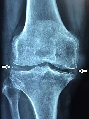
| X-ray of a knee with chondrocalcinosis | |
| Specialty | Radiology |
Chondrocalcinosis or cartilage calcification is calcification (accumulation of calcium salts) in hyaline cartilage and/or fibrocartilage.[1] It can be seen on radiography.
Signs and symptoms
The clinical presentation of chondrocalcinosis is as follows:[2]
- Chronic joint pain
- Swelling of joints
Causes
Buildup of calcium phosphate in the ankle joints has been found in about 50% of the general population, and may be associated with osteoarthritis.[3]
Another common cause of chondrocalcinosis is calcium pyrophosphate dihydrate crystal deposition disease (CPPD).[4] CPPD is estimated to affect 4% to 7% of the adult populations of Europe and the United States.[5] Previous studies have overestimated the prevalence by simply estimating the prevalence of chondrocalcinosis regardless of cause.[5]
A magnesium deficiency may cause chondrocalcinosis, and magnesium supplementation may reduce or alleviate symptoms.[6] In some cases, arthritis from injury can cause chondrocalcinosis.[7] Other causes of chondrocalcinosis include:[4]
- Hypercalcaemia, especially when caused by hyperparathyroidism
- Arthritis
- Pseudogout
- Wilson disease
- Hemochromatosis
- Ochronosis
- Hypophosphatasia
- Hypothyroidism
- Hyperoxalemia
- Acromegaly
- Gitelman syndrome
Diagnosis

Chondrocalcinosis can be visualized on projectional radiography, CT scan, MRI, US, and nuclear medicine.[1] CT scans and MRIs show calcific masses (usually within the ligamentum flavum or joint capsule), however radiography is more successful.[1] At ultrasound, chondrocalcinosis may be depicted as echogenic foci with no acoustic shadow within the hyaline cartilage.[8] As with most conditions, chondrocalcinosis can present with similarity to other diseases such as ankylosing spondylitis and gout.[1]
Treatment
The management of chondrocalcinosis is based on the following:[2]
References
- 1 2 3 4 Rothschild, Bruce M Calcium Pyrophosphate Deposition Disease (radiology)
- 1 2 "Chondrocalcinosis 2". NORD (National Organization for Rare Disorders). Archived from the original on 13 May 2022. Retrieved 22 September 2022.
- ↑ Hubert, Jan; Weiser, Lukas; Hischke, Sandra; Uhlig, Annemarie; Rolvien, Tim; Schmidt, Tobias; Butscheidt, Sebastian Karl; Püschel, Klaus; Lehmann, Wolfgang; Beil, Frank Timo; Hawellek, Thelonius (2018). "Cartilage calcification of the ankle joint is associated with osteoarthritis in the general population". BMC Musculoskeletal Disorders. 19 (1). doi:10.1186/s12891-018-2094-7. ISSN 1471-2474. PMC 5968601.
- 1 2 Matt A. Morgan; Frank Gaillard; et al. "Chondrocalcinosis". Radiopedia. Archived from the original on 2019-11-13. Retrieved 2017-08-11.
- 1 2 Ann K. Rosenthal. "Clinical manifestations and diagnosis of calcium pyrophosphate crystal deposition (CPPD) disease". UpToDate. Archived from the original on 2019-04-07. Retrieved 2022-03-15. This topic last updated: Jul 24, 2018.
- ↑ de Filippi JP, Diderich PP, Wouters JM (1992). "Hypomagnesemia and chondrocalcinosis". Ned Tijdschr Geneeskd. 136 (20): 139–41. PMID 1732847.
- ↑ Wright GD, Doherty M (1997). "Calcium pyrophosphate crystal deposition is not always 'wear and tear' or aging". Ann. Rheum. Dis. 56 (10): 586–8. doi:10.1136/ard.56.10.586. PMC 1752269. PMID 9389218.
- ↑ Arend CF. Ultrasound of the Shoulder. Master Medical Books, 2013. Free chapter on acromioclavicular chondrocalcinosis is available at ShoulderUS.com Archived 2017-07-14 at the Wayback Machine