Cruciate ligament of atlas
| Cruciate ligament of atlas | |
|---|---|
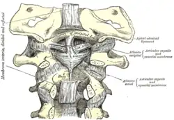 Membrana tectoria, transverse, and alar ligaments. ("Transverse ligament" and "vertical portion" visible intersecting at center.) | |
| Details | |
| System | skeletal |
| From | medial tubercles of atlas bone (C1), anterior side of foramen magnum of occipital bone of skull, body of axis bone (C2) |
| Identifiers | |
| Latin | ligamentum cruciforme atlantis |
| TA98 | A03.2.04.004 |
| TA2 | 1699 |
| FMA | 25018 |
| Anatomical terminology | |
The cruciate ligament of the atlas (cruciate may substitute for cruciform) is a ligament in the neck. It forms part of the atlanto-axial joint. The ligament is named after its cross shape. It consists of transverse and longitudinal components. The posterior longitudinal band may be absent in some people. The cruciate ligament of the atlas prevents abnormal movement of the atlanto-axial joint. It may be torn, such as by fractures of the atlas bone.

Structure
The cruciate ligament of the atlas consists of the transverse ligament of the atlas, a superior longitudinal band, and an inferior longitudinal band.[1][2] The superior longitudinal band connects the transverse ligament to the anterior side of the foramen magnum (near the basilar part) in the occipital bone of the skull.[1] The inferior longitudinal band connects the transverse ligament to the body of the axis bone (C2).[1]
Gerber's ligament runs deep to the superior band of the cruciate ligament of the atlas.[3]
Variation
The inferior longitudinal band may be absent in some people.[4] The rest of the ligament is always present.[4]
Function
The cruciate ligament of the atlas prevents abnormal movements of the atlanto-axial joint.[1] The longitudinal bands prevent hyperflexion and hyperextension of the occipital bone, and hold the transverse ligament of the atlas in a normal position.[1]
Clinical significance
Any part of the cruciate ligament of the atlas may tear, which is a significant injury. This may be caused by fractures of the atlas bone.[4] Ligament tears may be imaged with radiography, a CT scan, or magnetic resonance imaging.[4]
Ossification
Very rarely, the cruciate ligament of the atlas may ossify.[5] This may lead to cervical myelopathy, a deficit in the spinal cord.[5]
Etymology
The terms "cruciform" and "cruciate" refer to the cross shape of the ligament.[1] Both terms are frequently used, although the term "cruciate" may be confusing due to confusion with the anterior cruciate ligament and the posterior cruciate ligament of the knee.[4]
References
![]() This article incorporates text in the public domain from page 293 of the 20th edition of Gray's Anatomy (1918)
This article incorporates text in the public domain from page 293 of the 20th edition of Gray's Anatomy (1918)
- 1 2 3 4 5 6 Cramer, Gregory D. (2014). "5 - The Cervical Region". Clinical anatomy of the spine, spinal cord, and ANS (3rd ed.). St. Louis: Elsevier Health Sciences, Mosby. pp. 135–209. doi:10.1016/B978-0-323-07954-9.00005-0. ISBN 978-0-323-07954-9. OCLC 830314791.
- ↑ Federative Committee on Anatomical Terminology (1998). Terminologia anatomica: international anatomical terminology. Thieme. pp. 27–. ISBN 978-3-13-114361-7. Retrieved 17 June 2010.
- ↑ Ishak, Basem; Gnanadev, Raja; Dupont, Graham; Kikuta, Shogo; Altafulla, Juan; Iwanaga, Joe; Tubbs, R. Shane (2019-04-01). "Gerber's Ligament—A Forgotten Structure of the Craniocervical Junction". World Neurosurgery. Elsevier. 124: e707–e709. doi:10.1016/j.wneu.2018.12.198. ISSN 1878-8750. PMID 30660889. S2CID 58649895 – via ScienceDirect.
- 1 2 3 4 5 Tubbs, R. Shane; Iwanaga, Joe; Loukas, Marios; Kassem, Mohammad D. (2019-01-04). "The Cruciform Ligament". Clinical Anatomy of the Ligaments of the Craniocervical Junction. Cambridge Scholars Publishing. pp. 155–160. ISBN 978-1-5275-2418-7.
- 1 2 Baqai, Muhammad Waqas Saeed; Javed, Gohar; Baig, Mirza Zain (2019). "Ossification of the Cruciform Ligament of Atlas; a Rare Cause of Cervical Myelopathy: Case Report and Review of Literature". Asian Journal of Neurosurgery. 14 (3): 999–1003. doi:10.4103/ajns.AJNS_76_19. ISSN 1793-5482. PMC 6703042. PMID 31497151.
| Authority control: Scientific databases |
|---|