Eccrine angiomatous hamartoma
| Eccrine angiomatous hamartoma | |
|---|---|
 | |
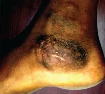
| Eccrine angiomatous hamartoma (left foot) | |
| Frequency | Lua error in Module:PrevalenceData at line 5: attempt to index field 'wikibase' (a nil value). |
Eccrine angiomatous hamartoma (EAH), first described by Lotzbeck in 1859, is a rare benign vascular hamartoma characterized histologically by a proliferation of eccrine and vascular components.[1][2] EAH exists on a spectrum of cutaneous tumors that include eccrine nevus, mucinous eccrine nevus and EAH. Each diagnostic subtype is characterized by an increase in the number as well as size of mature eccrine glands or ducts, with EAH being distinguished by the added vascular component.[3][4]
Patients with EAH may present with complaints of pain or increased sweating (hyperhidrosis) associated with stress or exercise, or without any associated symptoms.[3][4] It usually appears as a solitary nodular lesion on the acral areas of the extremities, particularly the palms and soles.[5] Onset of EAH most commonly arises in children prior to puberty as a solitary, unilateral, large, red to violaceous nodule or plaque located on the extremities.[3][4][6][7] Although rare, there have been reports of multiple EAH lesions occurring within a single patient in a linear, grouped, agminated or blaschkoid distribution.[8]
Signs and Symptoms
EAH most commonly presents as a solitary papule or plaque on the extremities of children and are frequently congenital, although they can appear in adulthood as well.[2][7] Rarely, multiple EAH lesions have been reported in a single patient, most often in an agminated pattern located on the extremities. A predisposition of EAH for facial and truncal involvement is not commonly seen. Some reports have demonstrated lesion predominance on the distal extremities.[6][7][9][10][11][12] Fewer accounts detail distribution on the head, neck and lower back.[3][8][13] Cases reporting lesions in uncommon locations, such as the trunk or abdomen, typically involved only a solitary lesion. Whereas EAH occurring as multiple lesions was more often reported in classic sites of involvement such as the arm or leg.[14][15]
Although EAH is often asymptomatic, it is known to cause variable levels of pain. This is thought to occur as a result of small nerves that are seen on electron microscopy in close proximity to the eccrine and vascular structures.[16] Hypertrichosis of the tumor is encountered in most cases. Hyperhidrosis is an additional diagnostic feature that is seen in under half of reported cases.[16] There is also significant cosmetic concern in some instances.[8][17]
Causes
The pathophysiologic mechanism underlying the hamartoma is thought to involve a biochemical fault in the interactions between differentiating epithelium and subjacent mesenchyme giving rise to an abnormal proliferation of adnexal and vascular structures.[2][17][18]
Diagnosis
A skin biopsy is typically performed for definitive diagnosis. The histopathologic hallmarks of EAH include the presence of an increased number of eccrine glands in the mid- and lower dermis along with ectatic or collapsed vessels that are seen in close approximation to the hyperplastic eccrine units. The overlying epidermis may be normal or may show acanthosis or papillomatosis.[3][4][6][16]
A recent report of EAH located on the neck described dermatoscopic features of multiple yellow, confluent nodules in a popcorn-like shape over a background of erythema and linear, arborizing vessels.[8] Dermoscopy is minimally invasive, inexpensive and may provide another diagnostic modality in the differentiation of EAH from other diagnoses, but has yet to be validated.[8]
Differential Diagnosis
Vascular malformations:
- Eccrine nevus – Characterized histopathologically by an increase in eccrine structures but not capillaries. Clinical hallmark is hyperhidrosis in most cases.
- Tufted angioma
- Smooth muscle hamartoma – These flesh-colored plaques may have associated hypertrichosis. A "chicken-skin" appearance (pseudo-Darier sign) may be seen with piloerection.
- Glomus tumor – Painful bluish papules, single or multiple, are encountered, mainly on acral areas of the body.
- Blue rubber bleb nevus
- Sudoriparous angioma – Another rare, benign tumor where eccrine glands of normal number are seen lying close to vascular structures in the dermis; these have a larger caliber than those seen in EAH.
Macules:
Treatment
EAH is a benign hamartoma and if there is no associated pain or cosmetic concern or disfiguration, EAH may be observed only. Treatment is often unnecessary.[7] Most cases of symptomatic EAH have been treated with surgical resection, with a few efficacious alternative treatments available.[19][20]
See also
References
- ↑ Lotzbeck (January 1859). "Ein Fall von Schweissdrüsengeschwulst an der Wange". Archiv für Pathologische Anatomie und Physiologie und für Klinische Medicin. 16 (1–2): 160–165. doi:10.1007/bf01945254. ISSN 0945-6317. Archived (PDF) from the original on 2021-08-29. Retrieved 2021-06-03.
- 1 2 3 García-Arpa, Mónica; Rodríguez-Vázquez, María; Cortina-de la Calle, Pilar; Romero-Aguilera, Guillermo; López-Pérez, Rafael (2005-01-01). "Multiple and Familial Eccrine Angiomatous Hamartoma". Acta Dermato-Venereologica. -1 (1): 355–7. doi:10.1080/00015550510027072. ISSN 0001-5555. PMID 16191863.
- 1 2 3 4 5 Tempark, T.; Shwayder, T. (2012-12-18). "Mucinous eccrine naevus: case report and review of the literature". Clinical and Experimental Dermatology. 38 (1): 1–6. doi:10.1111/ced.12034. ISSN 0307-6938. PMID 23252751.
- 1 2 3 4 Larralde, Margarita; Bazzolo, Eleonora; Boggio, Paula; Abad, María Eugenia; Santos Muñoz, Andrea (May 2009). "Eccrine Angiomatous Hamartoma: Report of Five Congenital Cases". Pediatric Dermatology. 26 (3): 316–319. doi:10.1111/j.1525-1470.2008.00777.x. ISSN 0736-8046. PMID 19706095.
- ↑ James, William D.; Elston, Dirk; Treat, James R.; Rosenbach, Misha A.; Neuhaus, Isaac (2020). "28. Dermal and subcutaneous tumors". Andrews' Diseases of the Skin: Clinical Dermatology (13th ed.). Edinburgh: Elsevier. p. 594. ISBN 978-0-323-54753-6. Archived from the original on 2022-10-05. Retrieved 2022-10-05.
- 1 2 3 Sulica, R. Lucien; Kao, Grace F.; Sulica, Virginia I.; Penneys, Neal S. (February 1994). "Eccrine angiomatous hamartoma (nevus):. Immunohistochemical findings and review of the literature". Journal of Cutaneous Pathology. 21 (1): 71–75. doi:10.1111/j.1600-0560.1994.tb00694.x. ISSN 0303-6987. PMID 7514619.
- 1 2 3 4 Nakatsui, Thomas C.; Schloss, Eric; Krol, Alfons; Lin, Andrew N. (July 1999). "Eccrine angiomatous hamartoma: Report of a case and literature review". Journal of the American Academy of Dermatology. 41 (1): 109–111. doi:10.1016/s0190-9622(99)70416-0. ISSN 0190-9622. PMID 10411421.
- 1 2 3 4 5 García-García, Sandra Cecilia; Saeb-Lima, Marcela; Villarreal-Martínez, Alejandra; Vázquez-Martínez, Osvaldo Tomás; López-Carrera, Yuri Igor; Ocampo-Candiani, Jorge; Gómez-Flores, Minerva (March 2018). "Dermoscopy of eccrine angiomatous hamartoma: The popcorn pattern". JAAD Case Reports. 4 (2): 165–167. doi:10.1016/j.jdcr.2017.08.014. ISSN 2352-5126. PMC 5789522. PMID 29387774.
- ↑ Zeller, D.J.; Goldman, R.L. (1971). "Eccrine-Pilar Angiomatous Hamartoma". Dermatology. 143 (2): 100–104. doi:10.1159/000252176. ISSN 1018-8665.
- ↑ Kikuchi, Ichiro; Kuroki, Yasumasa; Inoue, Shouhei (August 1982). "Painful Eccrine Angiomatous Nevus on the Sole". The Journal of Dermatology. 9 (4): 329–332. doi:10.1111/j.1346-8138.1982.tb02642.x. ISSN 0385-2407. PMID 6759552.
- ↑ Velasco, J.A.; Almeida, V. (1988). "Eccrine-Pilar Angiomatous Nevus". Dermatology. 177 (5): 317–322. doi:10.1159/000248587. ISSN 1018-8665. PMID 3072225.
- ↑ Sanmartin, Onofre; Botella, Rafael; Alegre, Victor; Martinez, Antonio; Aliaga, Adolfo (April 1992). "Congenital Eccrine Angiomatous Hamartoma". The American Journal of Dermatopathology. 14 (2): 161–164. doi:10.1097/00000372-199204000-00015. ISSN 0193-1091. PMID 1566976.
- ↑ Shin, Jaeyoung; Jang, Yong Hyun; Kim, Soo-Chan; Kim, You Chan (2013). "Eccrine Angiomatous Hamartoma: A Review of Ten Cases". Annals of Dermatology. 25 (2): 208–12. doi:10.5021/ad.2013.25.2.208. ISSN 1013-9087. PMC 3662915. PMID 23717013.
- ↑ Jorge-Finnigan, Conrado; Conejero, Claudia; Hernández-Martín, Angela; Sánchez-Gómez, Julian; Noguera-Morel, Lucero (March 2015). "Congenital Erythematous Plaques and Papules on the Right Arm". Pediatric Dermatology. 32 (2): 285–286. doi:10.1111/pde.12463. ISSN 0736-8046. PMID 25801080.
- ↑ CEBREIRO; SÁNCHEZ-AGUILAR; GÓMEZ CENTENO; FERNÁNDEZ-REDONDO; TORIBIO (November 1998). "Eccrine angiomatous hamartoma: report of seven cases". Clinical and Experimental Dermatology. 23 (6): 267–270. doi:10.1046/j.1365-2230.1998.00391.x. ISSN 0307-6938. PMID 10233623.
- 1 2 3 "VisualDx - Eccrine angiomatous hamartoma". VisualDx. Archived from the original on 2018-09-29. Retrieved 2018-09-29.
- 1 2 Pelle, Michelle T.; Pride, Howard B.; Tyler, William B. (September 2002). "Eccrine angiomatous hamartoma". Journal of the American Academy of Dermatology. 47 (3): 429–435. doi:10.1067/mjd.2002.121030. ISSN 0190-9622. PMID 12196755.
- ↑ Foshee, J. B.; Grau, Renee H.; Adelson, David M.; Crowson, Neil (July 2006). "Eccrine Angiomatous Harmartoma in an Infant". Pediatric Dermatology. 23 (4): 365–368. doi:10.1111/j.1525-1470.2006.00255.x. ISSN 0736-8046. PMID 16918635.
- ↑ Barco, Didac (2009-03-01). "Successful Treatment of Eccrine Angiomatous Hamartoma With Botulinum Toxin". Archives of Dermatology. 145 (3): 241–3. doi:10.1001/archdermatol.2008.575. ISSN 0003-987X. PMID 19289750.
- ↑ Felgueiras, João; del Pozo, Jesús; Sacristán, Felipe; Bonet, Maria del Mar (March 2015). "Eccrine Angiomatous Hamartoma". Dermatologic Surgery. 41 (3): 428–430. doi:10.1097/dss.0000000000000297. ISSN 1076-0512. PMID 25705946.