Great cardiac vein
| Great cardiac vein | |
|---|---|
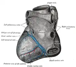
 Base and diaphragmatic surface of heart. (Great cardiac vein labeled at center left.) | |
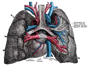
 Pulmonary vessels, seen in a dorsal view of the heart and lungs. The lungs have been pulled away from the median line, and a part of the right lung has been cut away to display the air-ducts and bloodvessels (great coronary vein labeled at center bottom). | |
| Details | |
| Drains to | Coronary sinus |
| Identifiers | |
| Latin | Vena cordis magna, vena cardiaca magna |
| TA98 | A12.3.01.003 |
| TA2 | 4159 |
| FMA | 4707 |
| Anatomical terminology | |
The great cardiac vein (left coronary vein) begins at the apex of the heart and ascends along the anterior longitudinal sulcus to the base of the ventricles.
It then curves around the left margin of the heart to reach the posterior surface. It merges with the oblique vein of the left atrium to form the coronary sinus,[1] which drains into the right atrium.
At the junction of the great cardiac vein and the coronary sinus, there is typically a valve present. This is the Vieussens valve of the coronary sinus.[1]
It receives tributaries from the left atrium and from both ventricles: one, the left marginal vein, is of considerable size, and ascends along the left margin of the heart.
References
![]() This article incorporates text in the public domain from page 642 of the 20th edition of Gray's Anatomy (1918)
This article incorporates text in the public domain from page 642 of the 20th edition of Gray's Anatomy (1918)
External links
- Anatomy photo:20:11-0101 at the SUNY Downstate Medical Center - "Heart: Cardiac veins"
- Anatomy figure: 20:03-05 at Human Anatomy Online, SUNY Downstate Medical Center - "Anterior view of the heart."