Maxillary veins
| Internal maxillary vein | |
|---|---|
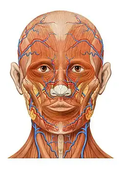
 Veins of the head and neck. (Internal maxillary vein visible at center.) | |
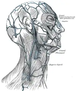
 Lateral head anatomy detail | |
| Details | |
| Drains to | retromandibular vein |
| Artery | maxillary artery |
| Identifiers | |
| Latin | venae maxillares |
| TA98 | A12.3.05.035 |
| TA2 | 4835 |
| FMA | 70850 |
| Anatomical terminology | |
The maxillary vein, or internal maxillary vein, is a vein of the head. It is a short trunk which accompanies the first part of the maxillary artery.
It is formed by a confluence of the veins of the pterygoid plexus and the interpterygoid emissary vein, and passes posteriorly between the sphenomandibular ligament and the neck of the mandible. It unites with the superficial temporal vein to form the retromandibular vein.
Structure
The maxillary vein is a short trunk which accompanies the first part of the maxillary artery. It is formed from the merging of the veins of the pterygoid plexus, and the interpterygoid emissary vein.[1] It passes posteriorly between the sphenomandibular ligament and the neck of the mandible. It unites with the superficial temporal vein.[2][3] It drains into the retromandibular vein (posterior facial vein).[2][3][4]
The maxillary vein anastomoses with the retroglenoid vein.[1]
Development
The maxillary vein may be the embryological origin of the central retinal vein.[5]
History
The maxillary vein may also be known as the internal maxillary vein.[1]
Other animals
The maxillary vein is found in many other mammals.[3]
Additional images
 Head anatomy anterior view
References
![]() This article incorporates text in the public domain from page 646 of the 20th edition of Gray's Anatomy (1918)
This article incorporates text in the public domain from page 646 of the 20th edition of Gray's Anatomy (1918)
- 1 2 3 Scremin, Oscar U. (2015). "31 - Cerebral Vascular System". The Rat Nervous System (4th ed.). Academic Press. pp. 985–1011. doi:10.1016/B978-0-12-374245-2.00031-0. ISBN 978-0-12-374245-2.
- 1 2 Thompson, Stevan H.; Yeung, Alison Y. (2016). "4 - Anatomy Relevant to Head, Neck, and Orofacial Infections". Head, Neck, and Orofacial Infections - A Multidisciplinary Approach. Elsevier Science. pp. 60–93. doi:10.1016/B978-0-323-28945-0.00004-1. ISBN 978-0-323-28945-0.
- 1 2 3 Cunningham Jr., Larry L.; Card, Aaron Sterling (2012). "38 - Mandibular Subcondylar Fractures". Current Therapy In Oral and Maxillofacial Surgery. Saunders. pp. 298–304. doi:10.1016/B978-1-4160-2527-6.00038-4. ISBN 978-1-4160-2527-6.
- ↑ Maynard, Robert Lewis; Downes, Noel (2019). "7 - The Cardiovascular System". Anatomy and Histology of the Laboratory Rat in Toxicology and Biomedical Research. Academic Press. pp. 77–90. doi:10.1016/B978-0-12-811837-5.00007-1. ISBN 978-0-12-811837-5.
- ↑ Remington, Lee Ann (2012). "7 - Ocular Embryology". Clinical Anatomy and Physiology of the Visual System (3rd ed.). Butterworth-Heinemann. pp. 123–143. doi:10.1016/B978-1-4377-1926-0.10007-4. ISBN 978-1-4377-1926-0.
| Authority control: Scientific databases |
|---|