Medial calcaneal branches of the tibial nerve
| Medial calcaneal branches of the tibial nerve | |
|---|---|
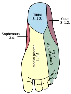 Diagram of the segmental distribution of the cutaneous nerves of the sole of the foot. | |
 Nerves of the right lower extremity Posterior view. (medial calcaneal labeled at bottom left.) Diagram of the segmental distribution of the cutaneous nerves of the sole of the foot. | |
| Details | |
| From | tibial nerve |
| Identifiers | |
| Latin | rami calcanei mediales nervi tibialis |
| TA98 | A14.2.07.065 |
| TA2 | 6589 |
| FMA | 44710 |
| Anatomical terms of neuroanatomy | |
The medial calcaneal branches of the tibial nerve (internal calcaneal branches) perforate the laciniate ligament, and supply the skin of the heel and medial side of the sole of the foot.[1]
Structure
The medial calcaneal nerve originates either from the tibial nerve or the lateral plantar nerve.[2] It splits into two cutaneous branches.[2]
Function
The medial calcaneal nerve provides sensory innervation to the medial side of the heel.[2]
See also
References
![]() This article incorporates text in the public domain from page 963 of the 20th edition of Gray's Anatomy (1918)
This article incorporates text in the public domain from page 963 of the 20th edition of Gray's Anatomy (1918)
- ↑ Charkhkar, Hamid; Shell, Courtney E; Marasco, Paul D; Pinault, Gilles J; Tyler, Dustin J; Triolo1, Ronald J (2018). "High-density peripheral nerve cuffs restore natural sensation to individuals with lower-limb amputations". Journal of Neural Engineering. 15 (5): 056002. doi:10.1088/1741-2552/aac964. PMID 29855427.
- 1 2 3 Ulcay, Tufan; Uzun, Ahmet; Ziylan, Taner (2014-09-01). "The origin and branching of medial calcaneal nerve in newborn foetuses". Journal of the Anatomical Society of India. 63: S1–S5. doi:10.1016/j.jasi.2014.06.001. ISSN 0003-2778 – via ScienceDirect.
| Authority control: Scientific databases |
|---|
This article is issued from Offline. The text is licensed under Creative Commons - Attribution - Sharealike. Additional terms may apply for the media files.