Positron emission mammography
| Positron emission mammography | |
|---|---|
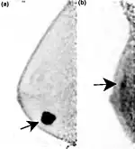
 Two PEM images, including sites of tracer uptake | |
| Purpose | imaging modality used to detect breast cancer |
Positron emission mammography (PEM) is a nuclear medicine imaging modality used to detect or characterise breast cancer.[1] Mammography typically refers to x-ray imaging of the breast, while PEM uses an injected positron emitting isotope and a dedicated scanner to locate breast tumors. Scintimammography is another nuclear medicine breast imaging technique, however it is performed using a gamma camera. Breasts can be imaged on standard whole-body PET scanners, however dedicated PEM scanners offer advantages including improved resolution.[2][3]
PEM is not recommended for routine use or for breast cancer screening, in part due to higher radiation dose compared to other modalities.[4] Compared to breast MRI, PEM offers higher specificity.[5] Specific indications can include "high-risk patients with masses > 2 cm or aggressive malignancy and serum tumor marker elevation".[6][7] 18F-FDG is the most common radiopharmaceutical used for PEM.[8]
Equipment
.png.webp)
PEM uses a specialised scanning system. Though some systems resemble a small PET scanner with a ring of detectors, others consist of a pair of gamma radiation detectors placed above and below the breast. On these systems, mild breast compression is applied to spread the breast and reduce its thickness. The detection process is identical to standard PET scanners. Positrons emitted by the injected 18F-FDG annihilate on interaction with electrons in tissue, leading to the emission of a pair of photons travelling in opposite directions. The detection of two simultaneous photons indicates the emission of a positron at a point on the line linking the two detection events. An image is the reconstructed from the collected emission data.[9][10]
History
Mammography using positron emitters was first proposed in 1994.[11] PEM is now approved in the United States and Europe for post-diagnosis imaging, with multiple commercial systems available.[12][13]
See also
References
- ↑ MacDonald, L.; Edwards, J.; Lewellen, T.; Haseley, D.; Rogers, J.; Kinahan, P. (October 2009). "Clinical imaging characteristics of the positron emission mammography camera: PEM Flex Solo II". J. Nucl. Med. 50 (10): 1666–75. doi:10.2967/jnumed.109.064345. PMC 2873041. PMID 19759118.
- ↑ Specht, Jennifer M; Mankoff, David A (16 March 2012). "Advances in molecular imaging for breast cancer detection and characterization". Breast Cancer Research. 14 (2): 206. doi:10.1186/bcr3094. PMC 3446362. PMID 22423895.
- ↑ Marino, Maria Adele; Helbich, Thomas H.; Blandino, Alfredo; Pinker, Katja (9 June 2015). "The role of positron emission tomography in breast cancer: a short review". Memo - Magazine of European Medical Oncology. 8 (2): 130–135. doi:10.1007/s12254-015-0210-z.
- ↑ Drukteinis, Jennifer S.; Mooney, Blaise P.; Flowers, Chris I.; Gatenby, Robert A. (June 2013). "Beyond Mammography: New Frontiers in Breast Cancer Screening". The American Journal of Medicine. 126 (6): 472–479. doi:10.1016/j.amjmed.2012.11.025. PMC 4010151. PMID 23561631.
- ↑ Kalles, Vasileios; Zografos, George C.; Provatopoulou, Xeni; Koulocheri, Dimitra; Gounaris, Antonia (13 December 2012). "The current status of positron emission mammography in breast cancer diagnosis". Breast Cancer. 20 (2): 123–130. doi:10.1007/s12282-012-0433-3. PMID 23239242.
- ↑ Fletcher, J. W.; Djulbegovic, B.; Soares, H. P.; Siegel, B. A.; Lowe, V. J.; Lyman, G. H.; Coleman, R. E.; Wahl, R.; Paschold, J. C.; Avril, N.; Einhorn, L. H.; Suh, W. W.; Samson, D.; Delbeke, D.; Gorman, M.; Shields, A. F. (20 February 2008). "Recommendations on the Use of 18F-FDG PET in Oncology". Journal of Nuclear Medicine. 49 (3): 480–508. doi:10.2967/jnumed.107.047787. PMID 18287273.
- ↑ Hosono, Makoto; Saga, Tsuneo; Ito, Kengo; Kumita, Shinichiro; Sasaki, Masayuki; Senda, Michio; Hatazawa, Jun; Watanabe, Hiroshi; Ito, Hiroshi; Kanaya, Shinichi; Kimura, Yuichi; Saji, Hideo; Jinnouchi, Seishi; Fukukita, Hiroyoshi; Murakami, Koji; Kinuya, Seigo; Yamazaki, Junichi; Uchiyama, Mayuki; Uno, Koichi; Kato, Katsuhiko; Kawano, Tsuyoshi; Kubota, Kazuo; Togawa, Takashi; Honda, Norinari; Maruno, Hirotaka; Yoshimura, Mana; Kawamoto, Masami; Ozawa, Yukihiko (31 May 2014). "Clinical practice guideline for dedicated breast PET". Annals of Nuclear Medicine. 28 (6): 597–602. doi:10.1007/s12149-014-0857-2. PMC 4328123. PMID 24878887.
- ↑ Cintolo, Jessica Anna; Tchou, Julia; Pryma, Daniel A. (16 March 2013). "Diagnostic and prognostic application of positron emission tomography in breast imaging: emerging uses and the role of PET in monitoring treatment response". Breast Cancer Research and Treatment. 138 (2): 331–346. doi:10.1007/s10549-013-2451-z. PMID 23504108.
- ↑ Glass and Shah (2013). "Clinical utility of positron emission mammography". Proc (Bayl Univ Med Cent). 26 (3): 314–9. doi:10.1080/08998280.2013.11928996. PMC 3684309. PMID 23814402.
- ↑ MacDonald, L.; Edwards, J.; Lewellen, T.; Haseley, D.; Rogers, J.; Kinahan, P. (16 September 2009). "Clinical Imaging Characteristics of the Positron Emission Mammography Camera: PEM Flex Solo II". Journal of Nuclear Medicine. 50 (10): 1666–1675. doi:10.2967/jnumed.109.064345. PMC 2873041. PMID 19759118.
- ↑ Thompson, C. J.; Murthy, K.; Weinberg, I. N.; Mako, F. (April 1994). "Feasibility study for positron emission mammography". Medical Physics. 21 (4): 529–538. Bibcode:1994MedPh..21..529T. doi:10.1118/1.597169. PMID 8058019.
- ↑ "Pros and Cons of Molecular Breast Imaging Tools". Imaging Technology News. 25 July 2012. Retrieved 5 January 2018.
- ↑ Subramaniam, Rathan (2015). PET/CT and Patient Outcomes, Part I, An Issue of PET Clinics. Elsevier Health Sciences. p. 161. ISBN 9780323370066.
![]() This article incorporates public domain material from the U.S. National Cancer Institute document: "Dictionary of Cancer Terms".
This article incorporates public domain material from the U.S. National Cancer Institute document: "Dictionary of Cancer Terms".