Primary cutaneous amyloidosis
| Primary cutaneous amyloidosis | |
|---|---|
| Other names: Primary localized cutaneous amyloidosis[1] | |
 | |
| Macular amyloidosis, located on the right lumbar region of the back | |
| Specialty | Dermatology |
Primary cutaneous amyloidosis is a form of amyloidosis associated with oncostatin M receptor.[2][3] This type of amyloidosis has been divided into the following types:[4]: 520
- Macular amyloidosis is a cutaneous condition characterized by itchy, brown, rippled macules usually located on the interscapular region of the back.[4]: 521 Combined cases of lichen and macular amyloidosis are termed biphasic amyloidosis, and provide support to the theory that these two variants of amyloidosis exist on the same disease spectrum.[5]
.jpg.webp) Macular amyloidosis
Macular amyloidosis.jpg.webp) Macular amyloidosis
Macular amyloidosis.jpg.webp) Macular amyloidosis
Macular amyloidosis
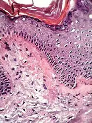
- Lichen amyloidosis is a cutaneous condition characterized by the appearance of occasionally itchy lichenoid papules, typically appearing bilaterally on the shins.[4]: 521
 Histopathology of lichen amyloidosis, with subepithelial Congo red-positive deposits
- Nodular amyloidosis is a rare cutaneous condition characterized by nodules that involve the acral areas.is a type of amyloidosis in skin.[6]
.jpg.webp)
Nodular amyloidosis
See also
References
- ↑ "Primary cutaneous amyloidosis | Genetic and Rare Diseases Information Center (GARD) – an NCATS Program". rarediseases.info.nih.gov. Archived from the original on 18 April 2019. Retrieved 18 April 2019.
- ↑ "Amyloid". Archived from the original on 2019-02-17. Retrieved 2021-04-17.
- ↑ Arita K, South AP, Hans-Filho G, et al. (January 2008). "Oncostatin M receptor-beta mutations underlie familial primary localized cutaneous amyloidosis". Am. J. Hum. Genet. 82 (1): 73–80. doi:10.1016/j.ajhg.2007.09.002. PMC 2253984. PMID 18179886.
- 1 2 3 James, William D.; Berger, Timothy G.; et al. (2006). Andrews' Diseases of the Skin: clinical Dermatology. Saunders Elsevier. ISBN 978-0-7216-2921-6.
- ↑ Lichen amyloidosis of the auricular concha Archived 2010-04-23 at the Wayback Machine Craig, E. (2006) Dermatology Online Journal 12 (5): 1, University of California, Davis Department of Dermatology
- ↑ Johnstone, Ronald B. (2017). "14. Cutaneous deposits". Weedon's Skin Pathology Essentials (2nd ed.). Elsevier. p. 286. ISBN 978-0-7020-6830-0. Archived from the original on 2021-05-25. Retrieved 2022-09-27.
External links
| Classification |
|---|
This article is issued from Offline. The text is licensed under Creative Commons - Attribution - Sharealike. Additional terms may apply for the media files.