Renal fascia
| Renal fascia | |
|---|---|
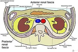
 Transverse plane through the kidneys, showing anterior and posterior renal fascia | |
| Details | |
| System | Urinary system |
| Identifiers | |
| Latin | fascia renalis |
| TA98 | A08.1.01.010 |
| TA2 | 3819 |
| FMA | 18104 |
| Anatomical terminology | |
The renal fascia is a layer of connective tissue encapsulating the kidneys and the adrenal glands. It can be divided into:
- The anterior renal fascia, also called Gerota's fascia (after Dimitrie Gerota)
- The posterior renal fascia, also called Zuckerkandl's fascia or fascia retrorenalis
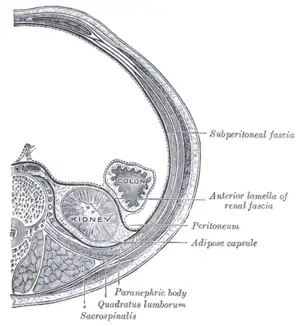
The renal fascia separates the adipose capsule of kidney from the overlying pararenal fat. The deeper layers below the renal fascia are, in order, the adipose capsule (or perirenal fat), the renal capsule and finally the parenchyma of the renal cortex.[1] The spaces about the kidney are typically divided into three compartments: the perinephric space and the anterior and posterior pararenal spaces.
Anterior renal fascia
- Anterior attachment: Passes anterior to the kidney, renal vessels, abdominal aorta and inferior vena cava and fuses with the anterior layer of the renal fascia of the opposite kidney.
- Posterior attachment: Fuses with the psoas fascia and side of the body of the vertebrae.
- Superior attachment: The anterior and posterior layers fuse at the upper pole of the kidney and then split to enclose the adrenal gland. At the upper part of the adrenal gland, they again fuse to form the suspensory ligament of the adrenal gland and fuse with the diaphragmatic fascia.
- Inferior attachment: The layers do not fuse. The posterior layer descends downwards and fuses with the iliac fascia. The anterior layer blends with the connective tissue of the iliac fossa.
The anterior fascia and posterior fascia fuse laterally to form the lateroconal fascia which fuses with the transverse fascia.[2]
In front of the fascia anterior to the perinephric space (also known as Gerota's fascia) is the anterior pararenal space, which contains the pancreas, ascending and descending colon, and second through fourth parts of the duodenum. The fascia posterior to the perinephric space was named Zuckerkandl's fascia. Posterior to this lies the posterior paranephric space which does not contain any abdominal organs.
Posterior renal fascia
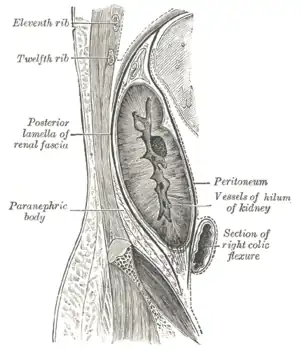
Zuckerkandl's fascia or fascia retrorenalis is a fibrous sheath covering the posterior side of the kidney. It constitutes the posterior layer of the renal fascia and was first described by Emil Zuckerkandl in 1883.[3]
Additional images
 Transverse section, showing the relations of the capsule of the kidney.
 Sagittal section through posterior abdominal wall, showing the relations of the capsule of the kidney.
References
![]() This article incorporates text in the public domain from page 1220 of the 20th edition of Gray's Anatomy (1918)
This article incorporates text in the public domain from page 1220 of the 20th edition of Gray's Anatomy (1918)
- ↑ Netter, Frank H. (2014). Atlas of Human Anatomy Including Student Consult Interactive Ancillaries and Guides (6th ed.). Philadelphia, Penn.: W B Saunders Co. p. 315. ISBN 978-1-4557-0418-7.
- ↑ Burkill, G J C.; Healy, J. C. (2000). "Anatomy of the retroperitoneum". Imaging. 12 (1): 10–20. doi:10.1259/img.12.1.120010.
- ↑ Chesbrough RM, Burkhard TK, Martinez AJ, Burks DD (1989). "Gerota versus Zuckerkandl: the renal fascia revisited". Radiology. 173 (3): 845–6. doi:10.1148/radiology.173.3.2682777. PMID 2682777.
External links
- Anatomy photo:40:03-0102 at the SUNY Downstate Medical Center - "Posterior Abdominal Wall: The Retroperitoneal Fat and Suprarenal Glands"
- Anatomy image:8951 at the SUNY Downstate Medical Center
- figures/chapter_29/29-5.HTM: Basic Human Anatomy at Dartmouth Medical School