Uterine prolapse
| Uterine prolapse | |
|---|---|
| Other names: Pelvic organ prolapse, prolapse of the uterus (womb), female genital prolapse, uterine descensus, uterine eversion | |
 | |
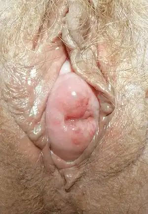
| Uterine prolapse in a 71-year-old woman, with the cervix visible in the vaginal opening. | |
| Specialty | Gynecology |
| Symptoms | Vaginal fullness, pain with sex, trouble urinating, urinary incontinence, constipation[1] |
| Usual onset | Gradual[2] |
| Types | 1st to 4th degree[1] |
| Risk factors | Pregnancy, childbirth, obesity, constipation, chronic cough[3] |
| Diagnostic method | Based on examination[1] |
| Differential diagnosis | Vaginal cancer, a long cervix[1][3] |
| Treatment | Pessary, hormone replacement therapy, surgery[1][3] |
| Frequency | About 14% of women[2] |
Uterine prolapse is when the uterus descends towards or through the opening of the vagina.[1] Symptoms may include vaginal fullness, pain with sex, trouble urinating, urinary incontinence, and constipation.[1] Often it gets worse over time.[2] Low back pain and vaginal bleeding may also occur.[3]
Risk factors include pregnancy, childbirth, obesity, constipation, and chronic coughing.[3] Diagnosis is based on examination.[1] It is a form of pelvic organ prolapse, together with bladder prolapse, large bowel prolapse, and small bowel prolapse.[4]
Preventive efforts include managing chronic breathing problems, not smoking, and maintaining a healthy weight.[3] Mild cases may be treated with a pessary together with hormone replacement therapy.[1][3] More severe cases may require surgery such as a vaginal hysterectomy.[1] About 14% of women are affected.[2] It occurs most commonly after menopause.[3][3]
Pathophysiology and causes
The uterus (womb) is normally held in place by a hammock of muscles and ligaments. Prolapse happens when the ligaments supporting the uterus become so weak that the uterus cannot stay in place and slips down from its normal position. These ligaments are the round ligament, uterosacral ligaments, broad ligament and the ovarian ligament. The uterosacral ligaments are by far the most important ligaments in preventing uterine prolapse.
In some cases of uterine prolapse, the uterus can be unsupported enough to extend past the vaginal wall for inches.[5]
The most common cause of uterine prolapse is trauma during childbirth, in particular multiple or difficult births. About 50% of women who have had children develop some form of pelvic organ prolapse in their lifetime.[6] It is more common as women get older, particularly in those who have gone through menopause. This condition is surgically correctable.
Treatment
Treatment is conservative, mechanical or surgical.
Conservative
Conservative options include behavioral modification and muscle strengthening exercises such as Kegel exercise.[7] Pessaries are a mechanical treatment as they elevate and support the uterus.[8][9]
Surgery
Surgical options are many[10] and may include a hysterectomy or a uterus-sparing technique such as laparoscopic hysteropexy,[11] sacrohysteropexy[12][13] or the Manchester operation.[14]
In the case of hysterectomy, the procedure can be accompanied by sacrocolpopexy.[15] This is a mesh-augmented procedure in which the apex of the vagina is attached to the sacrum by a piece of medical mesh material.[16]
A Cochrane review found that sacral colpopexy was associated with lower risk of complications than vaginal interventions, but it was unclear what route of sacral colpopexy should be preferred.[10] No clear conclusion could be reached regarding uterine preserving surgery versus vaginal hysterectomy for uterine prolapse. The evidence does not support use of transvaginal mesh (TVM) compared to native tissue repair for apical vaginal prolapse. The use of a transvaginal mesh is associated with side effects including pain, infection, and organ perforation. According to the FDA, serious complications are "not rare".
Society and culture
A number of class action lawsuits have been filed and settled against several manufacturers of TVM devices.
References
- 1 2 3 4 5 6 7 8 9 10 "Uterine and Vaginal Prolapse - Gynecology and Obstetrics". Merck Manuals Professional Edition. February 2017. Archived from the original on 15 October 2018. Retrieved 15 October 2018.
- 1 2 3 4 Culligan, Patrick J.; Goldberg, Roger P. (2007). Urogynecology in Primary Care. Springer Science & Business Media. p. 5. ISBN 9781846281679. Archived from the original on 15 October 2018. Retrieved 15 October 2018.
- 1 2 3 4 5 6 7 8 9 Ferri, Fred F. (2015). Ferri's Clinical Advisor 2016 E-Book: 5 Books in 1. Elsevier Health Sciences. p. 939. ISBN 9780323378222. Archived from the original on 27 August 2021. Retrieved 15 October 2018.
- ↑ "Uterine prolapse - Symptoms, diagnosis and treatment". BMJ Best Practice. Archived from the original on 15 October 2018. Retrieved 15 October 2018.
- ↑ D'Amico D, Barbarito C (10 February 2015). Health & physical assessment in nursing (3rd ed.). Boston. p. 665. ISBN 9780133876406. OCLC 894626609.
- ↑ "Oxford Gynaecological and Pelvic Floor Centre, Gynaecology in Oxford". www.oxfordgynaecology.com. Archived from the original on 25 April 2017. Retrieved 25 April 2017.
- ↑ Hagen, Suzanne (7 December 2011). "Conservative prevention and management of pelvic organ prolapse in women". The Cochrane Library (12): CD003882. doi:10.1002/14651858.CD003882.pub4. PMID 22161382.
- ↑ Bugge C, Adams EJ, Gopinath D, Reid F (February 2013). "Pessaries (mechanical devices) for pelvic organ prolapse in women" (PDF). Cochrane Database Syst Rev (2): CD004010. doi:10.1002/14651858.CD004010.pub3. PMID 23450548. Archived (PDF) from the original on 12 August 2017. Retrieved 26 October 2018.
- ↑ Cundiff GW, Amundsen CL, Bent AE, Coates KW, Schaffer JI, Strohbehn K, Handa VL (April 2007). "The PESSRI study: symptom relief outcomes of a randomized crossover trial of the ring and Gellhorn pessaries". Am. J. Obstet. Gynecol. 196 (4): 405.e1–8. doi:10.1016/j.ajog.2007.02.018. PMID 17403437. Archived (PDF) from the original on 27 August 2021. Retrieved 27 June 2019.
- 1 2 Maher, Christopher; Feiner, Benjamin; Baessler, Kaven; Christmann-Schmid, Corina; Haya, Nir; Brown, Julie (1 October 2016). "Surgery for women with apical vaginal prolapse" (PDF). The Cochrane Database of Systematic Reviews. 10: CD012376. doi:10.1002/14651858.CD012376. ISSN 1469-493X. PMID 27696355. Archived (PDF) from the original on 27 August 2021. Retrieved 27 June 2019.
- ↑ Rahmanou P, White B, Price N, Jackson S (January 2014). "Laparoscopic hysteropexy: 1- to 4-year follow-up of women postoperatively". Int Urogynecol J. 25 (1): 131–8. doi:10.1007/s00192-013-2209-5. PMID 24193261.
- ↑ Price N, Slack A, Jackson SR (January 2010). "Laparoscopic hysteropexy: the initial results of a uterine suspension procedure for uterovaginal prolapse". BJOG. 117 (1): 62–8. doi:10.1111/j.1471-0528.2009.02396.x. PMID 20002370.
- ↑ Rosati M, Bramante S, Conti F (August 2014). "A review on the role of laparoscopic sacrocervicopexy". Curr. Opin. Obstet. Gynecol. 26 (4): 281–9. doi:10.1097/GCO.0000000000000079. PMID 24950123.
- ↑ Surgical correction of uterine prolapse: cervical amputation with uterosacral ligament plication versus vaginal hysterectomy with high uterosacral ligament plication Archived 8 October 2011 at the Wayback Machine By de Boer T, Milani F, Kluivers K, Withagen M, Vierhout M. Part of ICS 2009 Scientific Programme, Thursday 1 October 2009
- ↑ Nygaard I, Brubaker L, Zyczynski HM, Cundiff G, Richter H, Gantz M, Fine P, Menefee S, Ridgeway B, Visco A, Warren LK, Zhang M, Meikle S (May 2013). "Long-term outcomes following abdominal sacrocolpopexy for pelvic organ prolapse". JAMA. 309 (19): 2016–24. doi:10.1001/jama.2013.4919. PMC 3747840. PMID 23677313.
- ↑ "Sacrocolpopexy with hysterectomy using mesh to repair uterine prolapse". NICE. GOV.UK. Archived from the original on 24 March 2018. Retrieved 28 March 2018.
External links
| Classification | |
|---|---|
| External resources |
- Illustrated description of Manchester Operation Archived 13 June 2017 at the Wayback Machine at atlasofpelvicsurgery.com
- Inverted uterus Archived 7 February 2018 at the Wayback Machine treatment, from Merck Professional