Subacromial bursa
| Subacromial bursa | |
|---|---|
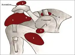
 Diagram of normal bursae surrounding the shoulder joint: (1) subacromial-subdeltoid bursa, (2) subscapular recess, (3) subcoracoid bursa, (4) coracoclavicular bursa, (5) supra-acromial bursa and (6) medial extension of subacromial-subdeltoid bursa. | |
| Details | |
| Identifiers | |
| Latin | bursa subacromialis |
| TA98 | A04.8.03.004 |
| TA2 | 2557 |
| FMA | 35515 35515, 35515 |
| Anatomical terminology | |
The subacromial bursa is the synovial cavity located just below the acromion, which communicates with the subdeltoid bursa in most individuals, forming the so-called subacromial-subdeltoid bursa (SSB).
The SSB bursa is located deep to the deltoid muscle and the coracoacromial arch and extends laterally beyond the humeral attachment of the rotator cuff, anteriorly to overlie the intertubercular groove, medially to the acromioclavicular joint, and posteriorly over the rotator cuff. The SSB decreases friction, and allows free motion of the rotator cuff relative to the coracoacromial arch and the deltoid muscle.
French anatomist and surgeon Jean-François Jarjavay is credited as the first to describe morbid processes of the SSB in 1867.[1] Since then, histologic studies have documented that synovial membrane may undergo inflammatory and/or degenerative changes and many now believe that they correspond to different stages in the spectrum of disease, with long-lasting inflammation leading to degeneration and fibrosis.[2]
See also
References
- ↑ Jarjavay JF. Sur la luxation du tendon de la longue portion du muscle biceps humeral: sur la luxation de tendons des muscles peroniers latercux. Gaz Hebd Med Chir 1867; 21:325.
- ↑ Arend CF. Ultrasound of the Shoulder. Master Medical Books, 2013. Read a sample chapter on the pathogenesis of shoulder bursitis at ShoulderUS.com
Sources
- Wheeless, Clifford. "Subacromial Bursa". Retrieved 2007-12-02.