Hobnail hemangioma
| Hobnail hemangioma | |
|---|---|
| Other names: Targetoid hemosiderotic hemangioma, superficial hemosiderotic lymphovascular malformation, targetoid hemosiderotic lymphovascular malformation[1] | |
 | |
| Specialty | Dermatology |
| Symptoms | Central brown or purplish small bump surrounded by bruise-like halo, typically on the trunk[2] |
| Usual onset | Young to middle-age adults[2] |
| Differential diagnosis | Melanoma[2] |
| Frequency | Rare, adults, males>females[1] |
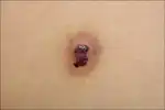
Hobnail hemangioma is a skin condition characterized by a central brown or purplish small bump that is surrounded by a bruise-like halo, typically on the trunk.[1][2]
It may appear similar to melanoma.[2]
It was first described by Santa Cruz and Aronberg in 1988.[2]
Signs and symptoms
 A dark brown papule with ecchymotic halo on left upper back
 Two dusky red to brown plaques with surrounding ecchymotic macular rings on the left knee
Diagnosis
 Wedge-shaped architecture and dilated vessels in the upper dermis (H&E, ×40)
 Intraluminal papillary projection and hobnail endothelial cells lining superficial dilated vessels (H&E, ×400)
See also
References
- 1 2 3 DE, Elder; D, Massi; RA, Scolyer; R, Willemze (2018). "Soft tissue tumours: Hobnail hemangioma". WHO Classification of Skin Tumours. Vol. 11 (4th ed.). Lyon (France): World Health Organization. pp. 347–348. ISBN 978-92-832-2440-2. Archived from the original on 2022-07-11. Retrieved 2022-08-08.
- 1 2 3 4 5 6 James, William D.; Elston, Dirk; Treat, James R.; Rosenbach, Misha A.; Neuhaus, Isaac (2020). "28. Dermal and subcutaneous tumors". Andrews' Diseases of the Skin: Clinical Dermatology (13th ed.). Edinburgh: Elsevier. p. 594. ISBN 978-0-323-54753-6. Archived from the original on 2022-10-05. Retrieved 2022-10-05.
This article is issued from Offline. The text is licensed under Creative Commons - Attribution - Sharealike. Additional terms may apply for the media files.