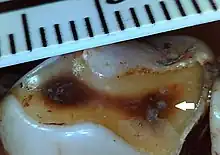
Tertiary dentin
Tertiary dentin (including reparative dentin or sclerotic dentin) forms as a reaction to stimulation, including caries, wear and fractures.[1] Tertiary dentin is therefore a mechanism for a tooth to ‘heal’, with new material formation protecting the pulp chamber and ultimately therefore protects the tooth and individual against abscesses and infection. This form of dentine can be easily distinguished on the surface of a tooth, and is much darker in appearance compared to primary dentine.[2] Tertiary dentine will often not be visible on the surface of a tooth, but because it is more dense it can be viewed on a Micro-CT scan of the tooth.[3]

Wear on the surface of a tooth can lead to the exposure of the underlying dentine. When wear is severe tertiary dentine may form to help protect the pulp chamber.[4] Frequency of tertiary dentin in different species of primate suggests teeth 'heal' at different rates in different species.[5] Gorillas have a high rate of tertiary dentin formation, with over 90% of worn teeth showing tertiary dentine.[5] Hominins have a much lower rate of tertiary dentin formation, with around 15% of teeth that have dentin exposed through wear showing tertiary dentin formation.[5] Chimpanzees have rates in between gorillas and humans, with 47% of worn teeth showing ‘healing’.
Clinical studies have researched the properties of tertiary dentine formation, including anatomy in both humans and animal models, usually from an oral health perspective.[6] Genetic changes in animal models can increase tertiary dentine production.[6] This suggests certain species may have evolved to produce tertiary dentin in response to dietary changes. For example, gorillas may have evolved high rates of tertiary dentin as protection against severe wear, since they consume a lot of tough vegetation.[1]
References
- 1 2 Towle, Ian (20 March 2019). "Tertiary Dentine Frequencies in Extant Great Apes and Fossil Hominins". Open Quaternary. 5 (1): 2. doi:10.5334/oq.48.
- ↑ Hillson, Simon (2005). Teeth. doi:10.1017/cbo9780511614477. ISBN 978-0-511-61447-7.
- ↑ Moggi-Cecchi, Jacopo; Menter, Colin; Dori, Irene; Irish, Joel D.; Riga, Alessandro; Towle, Ian (12 March 2019). "Root caries on a Paranthropus robustus third molar from Drimolen". bioRxiv: 573964. doi:10.1101/573964.
- ↑ Zuo, Jing; Zhen, Jiaxiu; Wang, Fei; Li, Yueheng; Zhou, Zhi (January 2018). "Effect of Low-Intensity Pulsed Ultrasound on the Expression of Calcium Ion Transport-Related Proteins during Tertiary Dentin Formation". Ultrasound in Medicine & Biology. 44 (1): 223–233. doi:10.1016/j.ultrasmedbio.2017.09.006. PMID 29079395.
- 1 2 3 Towle, Ian (20 March 2019). "Tertiary Dentine Frequencies in Extant Great Apes and Fossil Hominins". Open Quaternary. 5 (1): 2. doi:10.5334/oq.48.
- 1 2 Neves, V.C.M.; Sharpe, P.T. (29 November 2017). "Regulation of Reactionary Dentine Formation". Journal of Dental Research. 97 (4): 416–422. doi:10.1177/0022034517743431. PMID 29185832. S2CID 4027374.