Tornwaldt cyst
| Tornwaldt cyst | |
|---|---|
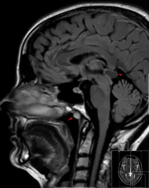 | |
| Tornwaldt cyst imaged on sagittal MRI (FLAIR). The cyst appears hyperintense in the midline of the nasopharynx (arrow). In this case there is also a cyst of the pinealis gland (arrowhead) showing a signal intensity slightly higher than the CSF. | |
| Specialty | ENT surgery |
A Tornwaldt cyst also spelt as Thornwaldt or Thornwald cyst[1] is a benign cyst located in the upper posterior nasopharynx. It can be seen on computed tomography (CT) or magnetic resonance imaging (MRI) of the head as a well-circumscribed round mass lying in the midline. In most cases, treatment is not necessary. It was first described by Gustav Ludwig Tornwaldt.
See also
References
- ↑ Gaillard, Frank. "Tornwaldt cyst | Radiology Reference Article | Radiopaedia.org". Radiopaedia. Retrieved 2019-10-02.
External links
| Wikimedia Commons has media related to Tornwaldt's cyst. |
This article is issued from Offline. The text is licensed under Creative Commons - Attribution - Sharealike. Additional terms may apply for the media files.