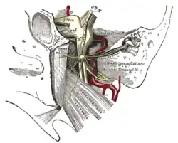
Trigeminal cave
| Trigeminal cave | |
|---|---|
 The trigeminal ganglion and its branches represented here as 1st division, 2nd division, and 3rd division. The Trigeminal Cave houses this ganglion. | |
| Details | |
| Identifiers | |
| Latin | cavum Meckeli, cavum trigeminale |
| TA98 | A14.1.01.108 |
| TA2 | 5379 |
| Anatomical terminology | |
The trigeminal cave (also known as Meckel's cave or cavum trigeminale) is a dura mater pouch containing cerebrospinal fluid.
Structure
The trigeminal cave is formed by the two layers of dura mater (endosteal and meningeal) which are part of an evagination of the cerebellar tentorium near the apex of the petrous part of the temporal bone. It envelops the trigeminal ganglion. It is bounded by the dura overlying four structures:
- cerebellar tentorium superolaterally
- lateral wall of the cavernous sinus superomedially
- clivus medially
- posterior petrous face inferolaterally
Within the dural confines of the trigeminal cave, there is a continuation of subarachnoid space along the posterior aspect of the cave, representing a continuation of the cerebral basal cisterns.[1]
History
Etymology
References
![]() This article incorporates text in the public domain from page 886 of the 20th edition of Gray's Anatomy (1918)
This article incorporates text in the public domain from page 886 of the 20th edition of Gray's Anatomy (1918)
- ↑ Burr HS, Robinson GB: An anatomical study of the gasserian ganglion with particular reference to the nature and extend of Meckel’s Cave (M,C). Anatomical Record 29:269-282, 1925.
- ↑ synd/2133 at Who Named It?
- ↑ J. F. Meckel. Tractatus anatomico physiologicus de quinto pare nervorum cerebri. Göttingen 1748.
This article is issued from Offline. The text is licensed under Creative Commons - Attribution - Sharealike. Additional terms may apply for the media files.