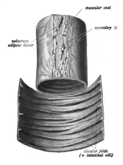
Circular folds
The circular folds (also known as valves of Kerckring, valves of Kerchkring, plicae circulares, plicae circulae, and valvulae conniventes) are large valvular flaps projecting into the lumen of the small intestine.
| Circular folds | |
|---|---|
 Small intestine (jejunus-ileum) with circular folds. | |
| Details | |
| Location | Small intestine |
| Identifiers | |
| Latin | plicae circulares |
| TA98 | A05.6.01.007 |
| TA2 | 2943 |
| FMA | 15071 |
| Anatomical terminology | |
Structure
The entire small intestine has circular folds of mucous membrane.[1] The majority extend transversely around the cylinder of the small intestine,[2] for about one-half or two-thirds of its circumference. Some form complete circles. Others have a spiral direction. The latter usually extend a little more than once around the bowel, but occasionally two or three times. While the larger folds are about 1 cm in depth at their broadest part, most folds are smaller. There tends to be an alternating pattern between larger and smaller folds.[1]
Distribution
They are not found at the commencement of the duodenum, but begin to appear about 2.5 or 5 cm beyond the pylorus.
In the lower part of the descending portion, below the point where the bile and pancreatic ducts enter the small intestine, they are very large and closely approximated.
In the horizontal and ascending portions of the duodenum and upper half of the jejunum they are large and numerous.[3] From this point, down to the middle of the ileum, they diminish considerably in size.
In the lower part of the ileum they almost entirely disappear; hence the comparative thinness of this portion of the intestine, as compared with the duodenum and jejunum.
Difference from other gastrointestinal folds
Unlike the gastric folds in the stomach, they are permanent, and are not obliterated when the intestine is distended.
The spaces between circular folds are smaller than the haustra of the colon, and, in contrast to haustra, circular folds reach around the whole circumference of the intestine. These differences can assist in distinguishing the small intestine from the colon on an abdominal x-ray.
Function
The circular folds slow the passage of the partly digested food along the intestines, and afford an increased surface for absorption.[4] They are covered with small finger-like projections called villi (singular, villus). Each villus, in turn, is covered with microvilli. The microvilli absorb fats and nutrients from the chyme.
History
The circular folds are also called the valves of Kerckring,[3] valves of Kerchkring,[5] plicae circulares,[3][4] plicae circulae,[5] and valvulae conniventes.[3]
References
![]() This article incorporates text in the public domain from page 1173 of the 20th edition of Gray's Anatomy (1918)
This article incorporates text in the public domain from page 1173 of the 20th edition of Gray's Anatomy (1918)
- Rumsey, D. (2005-01-01), "SMALL INTESTINE | Structure and Function", in Caballero, Benjamin (ed.), Encyclopedia of Human Nutrition (Second Edition), Oxford: Elsevier, pp. 126–133, doi:10.1016/b0-12-226694-3/02255-9, ISBN 978-0-12-226694-2, retrieved 2021-01-24
- Treuting, Piper M.; Arends, Mark J.; Dintzis, Suzanne M. (2018-01-01), Treuting, Piper M.; Dintzis, Suzanne M.; Montine, Kathleen S. (eds.), "11 - Upper Gastrointestinal Tract", Comparative Anatomy and Histology (Second Edition), San Diego: Academic Press, pp. 191–211, ISBN 978-0-12-802900-8, retrieved 2021-01-24
- Federle, Michael P.; Rosado-de-Christenson, Melissa L.; Raman, Siva P.; Carter, Brett W., eds. (2017-01-01), "Small Intestine", Imaging Anatomy: Chest, Abdomen, Pelvis (Second Edition), Elsevier, pp. 636–665, doi:10.1016/b978-0-323-47781-9.50031-3, ISBN 978-0-323-47781-9, retrieved 2021-01-24
- Yoder, Stephanie M.; Kindel, Tammy L.; Tso, Patrick (2010-01-01), Litwack, Gerald (ed.), "Chapter Eight - Using the Lymph Fistula Rat Model to Study Incretin Secretion", Vitamins & Hormones, Incretins and Insulin Secretion, Academic Press, 84: 221–249, doi:10.1016/B978-0-12-381517-0.00008-4, PMID 21094902, retrieved 2021-01-24
- Boudry, Gaëlle; Yang, Ping-Chang; Perdue, Mary H. (2004-01-01), "Small Intestine, Anatomy", in Johnson, Leonard R. (ed.), Encyclopedia of Gastroenterology, New York: Elsevier, pp. 404–409, doi:10.1016/b0-12-386860-2/00648-1, ISBN 978-0-12-386860-2, retrieved 2021-01-24
External links
- Anatomy photo:39:12-0302 at the SUNY Downstate Medical Center - "Intestines and Pancreas: The Jejunum and the Ileum"