Clostridium perfringens
Clostridium perfringens (formerly known as C. welchii, or Bacillus welchii) is a Gram-positive, bacillus (rod-shaped), anaerobic, spore-forming pathogenic bacterium of the genus Clostridium.[1][2] C. perfringens is ever-present in nature and can be found as a normal component of decaying vegetation, marine sediment, the intestinal tract of humans and other vertebrates, insects, and soil. It has the shortest reported generation time of any organism at 6.3 minutes in thioglycolate medium.[3]
| Clostridium perfringens | |
|---|---|
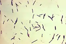 | |
| Photomicrograph of Gram-positive Clostridium perfringens bacilli | |
| Scientific classification | |
| Domain: | Bacteria |
| Phylum: | Bacillota |
| Class: | Clostridia |
| Order: | Eubacteriales |
| Family: | Lachnospiraceae |
| Genus: | Lachnoclostridium |
| Species: | C. perfringens |
| Binomial name | |
| Clostridium perfringens Veillon & Zuber 1898 Hauduroy et al. 1937 | |
Clostridium perfringens is one of the most common causes of food poisoning in the United States, alongside norovirus, Salmonella, Campylobacter, and Staphylococcus aureus.[4] However, it can sometimes be ingested and cause no harm.[5]
Infections due to C. perfringens show evidence of tissue necrosis, bacteremia, emphysematous cholecystitis, and gas gangrene, also known as clostridial myonecrosis. The specific name, perfringens, is derived from the Latin per (meaning "through") and frango ("burst"), referring to the disruption of tissue that occurs during gas gangrene.[6] The toxin involved in gas gangrene is α-toxin, which inserts into the plasma membrane of cells, producing gaps in the membrane that disrupt normal cellular function. C. perfringens can participate in polymicrobial anaerobic infections. It is commonly encountered in infections as a component of the normal flora. In this case, its role in disease is minor.
Tissue gas occurs when C. perfringens infects corpses. It causes extremely accelerated decomposition, and can only be stopped by embalming the corpse. Tissue gas most commonly occurs to those who have died from gangrene, large decubitus ulcers, necrotizing fasciitis or to those who had soil, feces, or water contaminated with C. perfrigens forced into an open wound.[7] These bacteria are resistant to the presence of formaldehyde in normal concentrations.
Genome
Clostridium perfringens has a stable G+C content around 27–28% and average genome size of 3.5 Mb.[8] Genomes of 56 C. perfringens strains have since been made available on NCBI genomes database for the scientific research community. Genomic research has revealed surprisingly high diversity in C. perfringens pangenome, with only 12.6% core genes, identified as the most divergent Gram-positive bacteria reported.[8] Nevertheless, 16S rRNA regions in between C. perfringens strains are found to be highly conserved (sequence identity >99.1%).[8]
Clostridium perfringens enterotoxin (CPE) gene
The clostridium perfrigens enterotoxin (CPE) producing strain has been identified to be a small portion of the overall C. perfringens population (~1-5%) through genomic testing.[9] Advances in gentic information surrounding strain A CPE C. perfringens has allowed techniques such as microbial source tracking (MST) to identify food contamination sources.[9] The CPE gene has been found within chromosomal DNA as well as plasmid DNA. Plasmid DNA has been shown to play and integral role in cell pathogenisis and encodes for major toxins, including CPE.[10]
Antibiotic Resistance
C. perfringens has been shown to carry plasmid containing genes for antibiotic resistance. The pCW3 plasmid is the primary conjugation plasmid responsible for creating antibiotic resistance in C. perfringens. Furthermore, the pCW3 plasmid also encodes for multiple toxins found in pathogenic strains of C. perfrigens.[11]
Motility
Although they lack flagella, C. perfringens bacteria are able to glide across surfaces because their bodies are lined with filaments from end-to-end. The hypermotile variants such as SM101, are often found arising on the edges of colonies on agar plates. Video microscopy of their gliding movement suggests that they form long, thin filaments that allow them to move rapidly like bacteria with flagella. Genome sequencing was used to identify the cause(s) of the hypermotile phenotype and their direct derivatives. In comparing them, strains SM124 and SM127, hypermotile derivatives of strains SM101 and SM102, respectively, contained 10 and six nucleotide polymorphisms (SNPs) relative to their parent strains. Mutations in cell division genes is the common feature of the hypermotile strains.[12]
Food poisoning
C. perfringens forms spores that are distributed through air, soil, and water. The most common cause of illness comes from the ingestion of poorly cooked meats that are contaminated by these spores.[13] After this meat is left out at 20 °C to 60 °C, the spores germinate and C. perfringens then grows rapidly. The bacteria produce a toxin that causes diarrhea.[14]
Food poisoning in humans is caused by type A strains able to produce C. perfringens enterotoxin.[15] This enterotoxin is a polypeptide of 35.5 kDa that accumulates in the beginning of the sporulation, and is excreted to the media when it lysates at the end of the sporulation. It is coded by the cpe gene, which is present in less than 5% of the type A strains, and it can be located in the chromosome or in an external plasmid[16]
In the United Kingdom and United States, C. perfringens bacteria are the third-most common cause of foodborne illness, with poorly prepared meat and poultry, or food properly prepared, but left to stand too long, the main culprits in harboring the bacterium.[17] The C. perfringens enterotoxin that mediates the disease is heat-labile (inactivated at 74 °C (165 °F)). It can be detected in contaminated food (if not heated properly), and feces.[18] Incubation time is between 6 and 25 (commonly 10–12) hours after ingestion of contaminated food.[19]
Since C. perfringens forms spores that can withstand cooking temperatures, if cooked food is left standing for long enough, germination can ensue and infective bacterial colonies develop. Symptoms typically include abdominal cramping, diarrhea, and fever.[20] The whole course usually resolves within 24 hours, but can last up to 2 weeks in older or infirm hosts.[21]
Clostridium perfringens poisoning can also lead to another disease known as enteritis necroticans or clostridial necrotizing enteritis, (also known as pigbel); this is caused by C. perfringens type C. This infection is often fatal. Large numbers of C. perfringens grow in the intestines, and secrete exotoxin. This exotoxin causes necrosis of the intestines, varying levels of hemorrhaging, and perforation of the intestine. Inflammation usually occurs in sections of the jejunum, midsection of the small intestine. This disease eventually leads to septic shock and death. This particular disease is rare in the United States; typically, it occurs in populations with a higher risk. Risk factors for enteritis necroticans include protein-deficient diet, unhygienic food preparation, sporadic feasts of meat (after long periods of a protein-deficient diet), diets containing large amounts of trypsin inhibitors (sweet potatoes), and areas prone to infection of the parasite Ascaris (produces a trypsin inhibitor). This disease is contracted in populations living in New Guinea, parts of Africa, Central America, South America, and Asia.[20]
Many cases of C. perfringens food poisoning likely remain subclinical, as antibodies to the toxin are common among the population. This has led to the conclusion that most of the population has experienced food poisoning due to C. perfringens.
Despite its potential dangers, C. perfringens is used as the leavening agent in salt-rising bread. The baking process is thought to reduce the bacterial contamination, precluding negative effects.[5]
Transmission and pathogenesis
C. perfringens is most commonly known for foodborne illness, but can translocate from a gastrointestinal source into the bloodstream which causes bacteremia. C. perfringens bacteremia can lead to toxin-mediated intravascular hemolysis and septic shock.[22] This is rare as it makes up less than 1% of bloodstream isolates, but is highly fatal with a reported mortality rate of 27% to 58%.[23]
Clostridium perfringens is the most common bacterial agent for gas gangrene. Some symptoms include blisters, tachycardia, swelling, and jaundice.[24]
A strain of C. perfringens might be implicated in multiple sclerosis (MS) nascent (Pattern III) lesions.[25] Tests in mice found that a two strains of intestinal C. perfringens that produced epsilon toxins (ETX) caused MS-like damage in the brain, and earlier work had identified this strain of C. perfringens in a human with MS.[26][27] MS patients were found to be 10 times more immune-reactive to the epsilon toxin than healthy people.[28]
Perfringolysin O (pfoA)-positive C. perfringens strains were also associated with the rapid onset of necrotizing enterocolitis in preterm infants.[29]
Major toxins
There are four major toxins produced by Clostridium perfringens. Alpha, beta, and epsilon toxins increase a cells permeability which causes an ion imbalance while iota toxins destroy the cell's actin cytoskeleton.[30] These toxins are produced by seven strains of C. perfringens, named A, B, C, D, E, F and G.[31]
Alpha toxin
Alpha toxin is a zinc-containing phospholipase C, composed of two structural domains, which destroy a cell's membrane. Alpha toxins are produced by all five types of C. perfringens. This toxin is linked to gas gangrene of humans and animals. Most cases of gas gangrene has been related to a deep wound being contaminated by soil that harbors C. perfringens.[30][32]
Beta toxin
Beta toxins are a protein that causes hemorrhagic necrotizing enteritis and enterotoxaemia in both animals (type B) and humans (type C) which leads to the infected individual's feces becoming bloody and their intestines necrotizing.[30]
Epsilon toxin
Epsilon toxin (ETX) is a protein produced by type B and type D strains of C. perfringens. This toxin is currently ranked the third most potent bacterial toxin known.[33] ETX causes enterotoxaemia in mainly goats and sheep, but cattle are sometime susceptible to it as well. A experiment using mice found that ETX had an LD50 of 50-110 ng/kg.[34] The excessive production of ETX increases the permeability of the intestines. This causes severe edema in organs such as the brain and kidneys.[35]
Iota toxin
Iota toxin is a protein produced by type E strains of C. perfringens. Iota toxins are made up of two, unlinked proteins that form a multimeric complex on cells. Iota toxins prevent the formation of filamentous actin. This causes the destruction of the cells cytoskeleton which in turn leads to the death of the cell as it can no longer maintain homeostasis.[36]
Diagnosis
Clostridium perfringens can be diagnosed by Nagler's reaction, in which the suspect organism is cultured on an egg yolk media plate. One side of the plate contains anti-alpha-toxin, while the other side does not. A streak of suspect organism is placed through both sides. An area of turbidity will form around the side that does not have the anti-alpha-toxin, indicating uninhibited lecithinase activity. Clostridium perfringens produces large colonies with irregular margins, often with a double zone of hemolysis.[37] In addition, laboratories can diagnose the bacteria by determining the number of bacteria in the feces. Within the 48 hours from when the disease began, if the individual has more than 106 spores of the bacteria per gram of stool, then the illness is diagnosed as C. perfringens food poisoning.[21]
Other tests/reactions: catalase – negative, spot indole – negative,[38] lecithinase – positive, lipase – negative, litmus milk – stormy fermentation; reverse CAMP plate – positive; gas liquid chromatography products – acetic, butyric and lactic acids.
Typically, the symptoms of C. perfringens poisoning are used to diagnose it. However, diagnosis can be made using a stool culture test, in which the feces are tested for toxins produced by the bacteria.[39]
Prevention
The growth of C. perfringens spores can be prevented by most cooking food, especially beef and poultry, to recommended temperatures. Leftover food should be refrigerated to a temperature below 40 °F (4 °C) within two hours of preparation. Large pots of food such as soup or stew with meats should be divided into small quantities and covered for refrigeration. Leftovers should be reheated to at least 165 °F (74 °C) before serving. A rule of thumb is that if the food tastes, smells, or looks different from what it is supposed to, then it should be avoided. Even if it looks safe, a food that has been out for a long time can also be dangerous to eat.[21]
The temperature that C. perfringens can multiply within can range anywhere from 59 °F (15 °C) to 122 °F (50 °C).[40]
Treatment
The most important aspect of treatment is prompt and extensive surgical debridement of the involved area and excision of all devitalized tissue, in which the organisms are prone to grow. Administration of antimicrobial drugs, particularly penicillin, is begun at the same time. Clostridium perfringens is more often susceptible to vancomycin compared to other pathogenic Clostridia.[41] Hyperbaric oxygen may be of help in the medical management of clostridial tissue infections.[42]
Most people who suffer from food poisoning caused by C. perfringens tend to fight off the illness without the need of any antibiotics. Extra fluids should be drank consistently until diarrhea dissipates.[43]
Epidemiology
Clostridium perfringens is a leading cause of food poisoning in the United States and Canada.[44] Contaminated meats in stews, soups, and gravies are usually responsible for outbreaks and cause about 1 million cases of foodborne illnesses in the United States every year.[43] Deaths due to the disease are rare and mostly occur in elderly and people who are predisposed to the disease.[45] From 1998 to 2010, 289 confirmed outbreaks of C. perfringens illness were reported with 15,208 illnesses, 82 hospitalizations, and eight deaths.[46]
Food poisoning incidents
On May 7, 2010, 42 residents and 12 staff members at a Louisiana (USA) state psychiatric hospital were affected and experienced vomiting, abdominal cramps, and diarrhea. Three patients died within 24 hours. The outbreak was linked to chicken which was cooked a day before it was served and was not cooled down according to hospital guidelines. The outbreak affected 31% of the residents of the hospital and 69% of the staff who ate the chicken. How many of the affected residents ate the chicken is unknown.[47]
In May 2011, a man died after allegedly eating food contaminated with the bacteria on a transatlantic American Airlines flight. The man's wife and daughter were suing American and LSG Sky Chefs, the German company that prepared the inflight food.[48]
In December 2012, a 46-year-old woman died two days after eating a Christmas Day meal at a pub in Hornchurch, Essex, England. She was among about 30 people to fall ill after eating the meal. Samples taken from the victims contained C. perfringens. The hotel manager and the cook were jailed for offences arising from the incident.[49]
In December 2014, 87-year-old Bessie Scott died three days after eating a church potluck supper in Nackawic, New Brunswick, Canada. Over 30 other people reported signs of gastrointestinal illness, diarrhea, and abdominal pain. The province's acting chief medical officer says, Clostridium perfringens is the bacteria [sic] that most likely caused the woman's death.[50]
In October 2016, 66-year-old Alex Zdravich died four days after eating an enchilada, burrito, and taco at Agave Azul in West Lafayette, Indiana, United States. Three others who dined the same day reported signs of foodborne illness, which were consistent with the symptoms and rapid onset of C. perfringens infection. They later tested positive for the presence of the bacteria, but the leftover food brought home by Zdravich tested negative.[51][52]
In November 2016, food contaminated with C. perfringens caused three individuals to die, and another 22 to be sickened, after a Thanksgiving luncheon hosted by a church in Antioch, California, United States.[53]
In January 2017, a mother and her son sued a restaurant in Rochester, New York, United States, as they and 260 other people were sickened after eating foods contaminated with C. perfringens. "Officials from the Monroe County Department of Public Health closed down the Golden Ponds after more than a fourth of its Thanksgiving Day guests became ill. An inspection revealed a walk-in refrigerator with food spills and mold, a damaged gasket preventing the door from closing, and mildew growing inside."[54]
In July 2018, 647 people reported symptoms after eating at a Chipotle Mexican Grill restaurant in Powell, Ohio, United States. Stool samples tested by the CDC tested positive for C. perfringens.[55]
In November 2018, approximately 300 people in Concord, North Carolina, United States, were sickened by food at a church barbecue that tested positive for C. perfringens.[56]
References
- Ryan, Kenneth J.; Ray, C. George (2004). Sherris Medical Microbiology : an Introduction to Infectious Diseases (4th ed.). New York: McGraw-Hill. p. 310. ISBN 978-0-8385-8529-0.
- Kiu, R; Hall, L. J. (2018). "An update on the human and animal enteric pathogen Clostridium perfringens". Emerging Microbes & Infections. 7 (141): 141. doi:10.1038/s41426-018-0144-8. PMC 6079034. PMID 30082713.
- "BioNumber Details Page". BioNumbers.
- "Foodborne Illnesses and Germs". Centers for Disease Control and Prevention (CDC). 2018-02-16. Retrieved 18 February 2018.
- Juckett, G; Bardwell, G; McClane, B; Brown, S (2008). "Microbiology of salt rising bread". The West Virginia Medical Journal. 104 (4): 26–7. PMID 18646681.
- Lexicon Orthopaedic Etymology. CRC Press. 1999. p. 128. ISBN 9789057025976.
- https://ldh.la.gov/assets/oph/Center-PHCH/Center-CH/infectious-epi/EpiManual/ClostridiumPerfringensManual.pdf
- Kiu, Raymond; Caim, Shabhonam; Alexander, Sarah; Pachori, Purnima; Hall, Lindsay J. (2017). "Probing Genomic Aspects of the Multi-Host Pathogen Clostridium perfringens Reveals Significant Pangenome Diversity, and a Diverse Array of Virulence Factors". Frontiers in Microbiology. 8: 2485. doi:10.3389/fmicb.2017.02485. PMC 5733095. PMID 29312194.
- Miyamoto, Kazuaki; Li, Jihong; McClane, Bruce A. (2012). "Enterotoxigenic Clostridium perfringens: Detection and Identification". Microbes and environments. 27 (4): 343–349. doi:10.1264/jsme2.ME12002. ISSN 1342-6311.
- Gulliver, Emily L.; Adams, Vicki; Marcelino, Vanessa Rossetto; Gould, Jodee; Rutten, Emily L.; Powell, David R.; Young, Remy B.; D’Adamo, Gemma L.; Hemphill, Jamia; Solari, Sean M.; Revitt-Mills, Sarah A.; Munn, Samantha; Jirapanjawat, Thanavit; Greening, Chris; Boer, Jennifer C. (2023-04-20). "Extensive genome analysis identifies novel plasmid families in Clostridium perfringens". Microbial Genomics. 9 (4). doi:10.1099/mgen.0.000995. ISSN 2057-5858.
- Adams, Vicki; Han, Xiaoyan; Lyras, Dena; Rood, Julian I. (September 2018). "Antibiotic resistance plasmids and mobile genetic elements of Clostridium perfringens". Plasmid. 99: 32–39. doi:10.1016/j.plasmid.2018.07.002.
- Liu, Hualan; McCord, Kristin D.; Howarth, Jonathon; David, Popham L.; Jensen, Roderick V.; Melville, Stephen B. (July 2014). "Hypermotility in Clostridium perfringens Strain SM101 Is Due to Spontaneous Mutations in Genes Linked to Cell Division". Journal of Bacteriology. 196 (13): 2405–2412. doi:10.1128/JB.01614-14. PMC 4054169. PMID 24748614.
- "Prevent Illness From C. perfringens", CDC
- "Clostridium perfringens Food Poisoning - Infectious Diseases". Merck Manuals Professional Edition. Retrieved 2023-10-17.
- Kiu, Raymond; Caim, Shabhonam; Painset, Anais; Pickard, Derek; Swift, Craig; Dougan, Gordon; Mather, Alison E.; Amar, Corinne; Hall, Lindsay J. (25 September 2019). "Phylogenomic analysis of gastroenteritis-associated Clostridium perfringens in England and Wales over a 7-year period indicates distribution of clonal toxigenic strains in multiple outbreaks and extensive involvement of enterotoxin-encoding (CPE) plasmids". Microbial Genomics. 5 (10). doi:10.1099/mgen.0.000297. PMC 6861862. PMID 31553300.
- Labbe, R.G.; Juneja, V.K. (2017), "Clostridium perfringens", Foodborne Diseases, Elsevier, pp. 235–242, doi:10.1016/b978-0-12-385007-2.00010-3
- Warrell; et al. (2003). Oxford Textbook of Medicine (4th ed.). Oxford University Press. ISBN 978-0-19-262922-7.
- Murray; et al. (2009). Medical Microbiology (6th ed.). Mosby Elsevier. ISBN 978-0-323-05470-6.
- Fafangel, Mario; UČacar, Veribuca; Vudrag, Marko; Berce, Ingrid; Kraigher, Alenka (2015). "A Five Site Clostridium Perfringens Food-Borne Outbreak: A Retrospective Cohort Study". Slovenian Journal of Public Health. 54 (1): 51–57. doi:10.1515/sjph-2015-0007. PMC 4820149. PMID 27646622.
- Lentino, Joseph R. (2016-01-01). "Clostridial Necrotizing Enteritis". Merck Manuel. Merck Sharp & Dohme Corp. Retrieved 2016-09-27.
- "Clostridium perfringens". Center for Disease Control and Prevention. 2015-10-08. Retrieved 2016-09-27.
- "Cytarabine". Reactions Weekly. 1959 (1): 223–223. 2023-06-03. doi:10.1007/s40278-023-40395-7. ISSN 1179-2051.
- Millard, Michael A.; McManus, Kathleen A.; Wispelwey, Brian (2016). "Severe Sepsis due to Clostridium perfringens Bacteremia of Urinary Origin: A Case Report and Systematic Review". Case Reports in Infectious Diseases. 2016: 1–5. doi:10.1155/2016/2981729. ISSN 2090-6625.
- "Gas gangrene: MedlinePlus Medical Encyclopedia".
- Rumah, Kareem Rashid; Linden, Jennifer; Fischetti, Vincent A.; Vartanian, Timothy; Esteban, Francisco J. (16 October 2013). "Isolation of Clostridium perfringens Type B in an Individual at First Clinical Presentation of Multiple Sclerosis Provides Clues for Environmental Triggers of the Disease". PLOS ONE. 8 (10): e76359. Bibcode:2013PLoSO...876359R. doi:10.1371/journal.pone.0076359. PMC 3797790. PMID 24146858.
- "Multiple sclerosis 'linked to food bug'". BBC. 29 January 2014. Retrieved 29 January 2014.
- Reder, Anthony T. (2023-05-01). "Clostridium epsilon toxin is excessive in multiple sclerosis and provokes multifocal lesions in mouse models". Journal of Clinical Investigation. 133 (9). doi:10.1172/JCI169643. ISSN 1558-8238.
- Woerner, Amanda (29 January 2014). "Bacterial toxin may trigger multiple sclerosis, research finds". Fox News.
- Kiu, Raymond; Shaw, Alexander G.; Sim, Kathleen; Acuna-Gonzalez, Antia; Price, Christopher A.; Bedwell, Harley; Dreger, Sally A.; Fowler, Wesley J.; Cornwell, Emma; Pickard, Derek; Belteki, Gusztav; Malsom, Jennifer; Phillips, Sarah; Young, Gregory R.; Schofield, Zoe (June 2023). "Particular genomic and virulence traits associated with preterm infant-derived toxigenic Clostridium perfringens strains". Nature Microbiology. 8 (6): 1160–1175. doi:10.1038/s41564-023-01385-z. ISSN 2058-5276. PMC 10234813. PMID 37231089.
- Stiles, Bradley G.; Barth, Gillian; Barth, Holger; Popoff, Michel R. (2013-11-12). "Clostridium perfringens Epsilon Toxin: A Malevolent Molecule for Animals and Man?". Toxins. 5 (11): 2138–2160. doi:10.3390/toxins5112138. ISSN 2072-6651. PMC 3847718. PMID 24284826.
- Johnston MD, Whiteside TE, Allen ME, Kurtz DM (February 2022). "Toxigenic Profile of Clostridium perfringens Strains Isolated from Natural Ingredient Laboratory Animal Diets". Comparative Medicine. 72 (1): 50–58. doi:10.30802/AALAS-CM-22-000013. PMC 8915413. PMID 35148812.
- Li, Ming; Li, Ning (2021-06-16). "Clostridium perfringens bloodstream infection secondary to acute pancreatitis: A case report". World Journal of Clinical Cases. 9 (17): 4357–4364. doi:10.12998/wjcc.v9.i17.4357. ISSN 2307-8960.
- Alves, Guilherme Guerra; Machado de Ávila, Ricardo Andrez; Chávez-Olórtegui, Carlos Delfin; Lobato, Francisco Carlos Faria (2014-12-01). "Clostridium perfringens epsilon toxin: The third most potent bacterial toxin known". Anaerobe. 30: 102–107. doi:10.1016/j.anaerobe.2014.08.016. ISSN 1075-9964.
- Xin, Wenwen; Wang, Jinglin (2019-09-01). "Clostridium perfringens epsilon toxin: Toxic effects and mechanisms of action". Biosafety and Health. 1 (2): 71–75. doi:10.1016/j.bsheal.2019.09.004. ISSN 2590-0536.
- Geng, Zhijun; Kang, Lin; Huang, Jing; Gao, Shan; Wang, Jing; Yuan, Yuan; Li, Yanwei; Wang, Jinglin; Xin, Wenwen (2021-07-30). "Epsilon toxin from Clostridium perfringens induces toxic effects on skin tissues and HaCaT and human epidermal keratinocytes". Toxicon. 198: 102–110. doi:10.1016/j.toxicon.2021.05.002. ISSN 0041-0101.
- Sakurai, Jun; Nagahama, Masahiro; Oda, Masataka; Tsuge, Hideaki; Kobayashi, Keiko (2009-12-23). "Clostridium perfringens Iota-Toxin: Structure and Function". Toxins. 1 (2): 208–228. doi:10.3390/toxins1020208. ISSN 2072-6651. PMC 3202787. PMID 22069542.
- "Clostridium perfringens" (PDF). Louisiana Department of Health. 2015-11-24. Retrieved 2023-03-06.
- Harpold, D. J.; Wasilauskas, B. L.; O'Connor, M. L. (1985). "Rapid identification of Clostridium species by high-pressure liquid chromatography" (PDF). Journal of Clinical Microbiology. 22 (6): 962–967. doi:10.1128/jcm.22.6.962-967.1985. PMC 271860. PMID 3905852.
- "Bad Bug Book (Second Edition)" (PDF). U.S. Food and Drug Administration. 2014-10-07. Retrieved 2016-09-26.
- Taormina, Peter J.; Dorsa, Warren J. (July 2004). "Growth Potential of Clostridium perfringens during Cooling of Cooked Meats". Journal of Food Protection. 67 (7): 1537–1547. doi:10.4315/0362-028X-67.7.1537.
- Di Bella, Stefano; Antonello, Roberta Maria; Sanson, Gianfranco; Maraolo, Alberto Enrico; Giacobbe, Daniele Roberto; Sepulcri, Chiara; Ambretti, Simone; Aschbacher, Richard; Bartolini, Laura; Bernardo, Mariano; Bielli, Alessandra (June 2022). "Anaerobic bloodstream infections in Italy (ITANAEROBY): A 5-year retrospective nationwide survey". Anaerobe. 75: 102583. doi:10.1016/j.anaerobe.2022.102583. hdl:11368/3020691. PMID 35568274. S2CID 248736289.
- Jawetz Melnick & Adelbergs Medical Microbiology - 27E.
- CDC (2023-03-24). "Prevent Illness From C. perfringens". Centers for Disease Control and Prevention. Retrieved 2023-10-01.
- Johnson, E. A., Summanen, P., & Finegold, S. M. (2007). Clostridium. In P. R. Murray (Ed.), Manual of Clinical Microbiology (9th ed., pp. 889-910). Washington, D.C.: ASM Press
- Songer, J. G. (2010). Clostridia as agents of zoonotic disease. Veterinary Microbiology, 140(3-4), 399-404
- Grass, Julian E.; Gould, L. Hannah; Mahon, Barbara E. (1 February 2013). "Epidemiology of foodborne disease outbreaks caused by Clostridium perfringens, United States, 1998-2010". Foodborne Pathog. Dis. 10 (2): 131–136. doi:10.1089/fpd.2012.1316. PMC 4595929. PMID 23379281.
- "Fatal Foodborne Clostridium perfringens Illness at a State Psychiatric Hospital — Louisiana, 2010". Centers for Disease Control and Prevention. Retrieved 16 November 2013.
- Mohn, Tanya (1 December 2011). "Passenger dies in-flight, family says airline to blame". Overhead Bin. MSNBC. Retrieved 2012-02-13.
- "Pub chef and manager jailed over Christmas dinner that left a diner dead". The Guardian. 23 January 2015. Retrieved 3 August 2015.
- "Woman's death likely caused by bacteria from Christmas supper". CBC. 12 December 2014.
- "Food poisoning death at Indiana restaurant kept secret for months". 13 WTHR Indianapolis. 2017-07-17. Retrieved 2017-07-18.
- (WTHR), Susan Batt. "Agave Azul Tippecanoe Co Food Poisoning Finding Summary". www.documentcloud.org. Retrieved 2017-07-18.
- "Bacteria that killed 3 at Antioch Thanksgiving dinner pinpointed". SFGate. Retrieved 2016-12-20.
- "Mother, son sue eatery for Thanksgiving dinner food poisoning - Food Safety News". 6 January 2017.
- "CDC releases test findings after hundreds sickened at Powell Chipotle - Columbus Dispatch". 16 August 2018.
- "Strain of food poisoning causes illness at North Carolina church barbecue". November 2018.
External links
- Clostridium perfringens genomes and related information at PATRIC, a Bioinformatics Resource Center funded by NIAID
- Pathema-Clostridium Resource
- Type strain of Clostridium perfringens at BacDive - the Bacterial Diversity Metadatabase
| Adulterants, food contaminants | |
|---|---|
| Food additives | |
| Intestinal parasites and parasitic disease | |
| Microorganisms | |
| Pesticides | |
| Preservatives | |
| Sugar substitutes | |
| Toxins, poisons, environment pollution | |
| Food processing | |
| Food contamination incidents |
|
| Regulation, standards, watchdogs | |
| Institutions |
|
| Related topics | |
| |