Dolichousnea
Dolichousnea is a genus of fruticose lichens in the family Parmeliaceae. It has three species.[3] The widely distributed type species, Dolichousnea longissima, is found in boreal regions of Asia, Europe, and North America.
| Dolichousnea | |
|---|---|
 | |
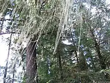
| Dolichousnea longissima in a coniferous forest in Washington (state), USA | |
| Scientific classification | |
| Domain: | Eukaryota |
| Kingdom: | Fungi |
| Division: | Ascomycota |
| Class: | Lecanoromycetes |
| Order: | Lecanorales |
| Family: | Parmeliaceae |
| Genus: | Dolichousnea (Y.Ohmura) Articus (2004)[1] |
| Type species | |
| Usnea longissima Ach. (1810) | |
| Species | |
|
D. diffracta | |
| Synonyms[2] | |
| |
Taxonomy
Dolichousnea was originally circumscribed as a subgenus of the large genus Usnea, by Japanese lichenologist Yoshihito Ohmura in 2001. He considered that it was possible to treat the three taxa as a genus, but thought it more suitable to treat Dolichousnea at the rank of subgenus, because the taxa have the synapomorphies of the genus Usnea: a fruticose thallus, a central cartilaginous axis throughout the thallus, and the presence of usnic acid in the cortex.[4] In 2004, Kristina Articus proposed to raise the subgenus to generic rank,[1] a decision that was accepted in later analyses.[5][3]
Description
Dolichousnea lichens have a pendent thallus, in which all the branches hang downward. The branching is isotomic-dichotomous, meaning that the thallus divides into isotomic (i.e. dividing a line at points equally distant from its opposite ends) branches of more or less equal thickness. One of the characteristics of Dolichousnea are the presence of annular-pseudocyphellae between segments.[6] These are pseudocyphellae that resemble annular cracks, but the space between segments is swollen and surrounded by maculae (white spots on branch surface that form when the cortex tissue is thin).[7] These pseudocyphellae differ from physical cracks, and seem to be formed due to pressure from the medullary hyphae beneath the cortex.[8] Dolichousnea has a thicker hypothecium (a layer of dense hyphal tissue just below the hymenium) than other subgenera of Usnea, typically measuring 100–160 μm; in contrast, subgenera Usnea and Eumitria range over 30–60 μm.[9] Another characteristic of Dolichousnea is a positive iodine reaction in its axis.[10]
Species
- Dolichousnea diffracta (Vain.) Articus (2004) – East Asia
- Dolichousnea longissima (Ach.) Articus (2004) – boreal regions of Asia, Europe, North America
- Dolichousnea trichodeoides (Vain. ex Motyka) Articus (2004) – East Africa; Asia; Australia; Europe
Recent (2020) molecular phylogenetic analysis suggests that the taxon Dolichousnea trichodeoides represent an unresolved species complex.[11]
References
- Articus, Kristina (2004). "Neuropogon and the phylogeny of Usnea s.l. (Parmeliaceae, Lichenized Ascomycetes)". Taxon. 53 (4): 925–934. doi:10.2307/4135560. JSTOR 4135560.
- "Synonymy: Dolichousnea (Y. Ohmura) Articus, Taxon 53(4): 932 (2004)". Species Fungorum. Retrieved 17 March 2021.
- Wijayawardene, Nalin; Hyde, Kevin; Al-Ani, Laith Khalil Tawfeeq; Somayeh, Dolatabadi; Stadler, Marc; Haelewaters, Danny; et al. (2020). "Outline of Fungi and fungus-like taxa". Mycosphere. 11: 1060–1456. doi:10.5943/mycosphere/11/1/8.
- Ohmura 2001, p. 80.
- Divakar, Pradeep K.; Crespo, Ana; Kraichak, Ekaphan; Leavitt, Steven D.; Singh, Garima; Schmitt, Imke; Lumbsch, H. Thorsten (2017). "Using a temporal phylogenetic method to harmonize family- and genus-level classification in the largest clade of lichen-forming fungi". Fungal Diversity. 84 (1): 101–117. doi:10.1007/s13225-017-0379-z. S2CID 256071368.
- Ohmura 2001, p. 8.
- Ohmura 2001, p. 9.
- Ohmura 2001.
- Ohmura 2001, p. 17.
- Ohmura 2001, p. 21.
- Lücking, Robert; Nadel, Miko; Araujo, Elena; Gerlach, Alice (2020). "Two decades of DNA barcoding in the genus Usnea (Parmeliaceae): how useful and reliable is the ITS?". Plant and Fungal Systematics. 65 (2): 303–357. doi:10.35535/pfsyst-2020-0025.
Cited literature
- Ohmura, Y. (2001). "Taxonomic study of the genus Usnea (lichenized Ascomycetes) in Japan and Taiwan". Journal of the Hattori Botanical Laboratory. 90: 1–96.