Dural tail sign
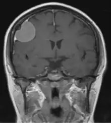
The dural tail sign (also known as "dural thickening", "flare sign", or "meningeal sign") is a radiological finding observed in magnetic resonance imaging (MRI) studies of the brain that refers to a thickening of the dura mater immediately adjacent to a mass lesion, such as a brain tumor.[1] Initially, the dural tail sign was thought to be pathognomonic of meningioma, a slow-growing tumor that arises from the meninges.[1] However, subsequent studies have shown that it can also be observed in various intra- and extra-cranial pathologies and in spinal lesions.[1] It is not a completely sensitive finding, as it is seen in only 60-72% of cases.[2] It is not completely specific either, as it has been described associated with lesions like neuromas, chloromas, pituitary diseases, granulomatous disorders, cerebral Erdheim-Chester disease, lymphomas, metastasis, hemangiopericytomas, schwannomas, and gliomas such as glioblastoma multiforme (GBM).[2][3] The final diagnosis should be further established through cerebrospinal fluid analysis or histopathological examination following a biopsy.[3]
The dural tail sign was first described in 1989 by Wilms et al..[1][4] Histopathological correlation from different studies has at times revealed tumor infiltration into the dura mater, however, in most instances, it signifies a hypervascular, non-neoplastic response.[3]
One study showed that including the dural tail in the stereotactic radiosurgery (SRS) or fractionated stereotactic radiotherapy (FSRT) volumes for meningioma treatment did not seem to impact recurrence.[5]
References
- Sotoudeh, Houman (2010). "A review on dural tail sign". World Journal of Radiology. Baishideng Publishing Group Inc. 2 (5): 188–192. doi:10.4329/wjr.v2.i5.188. ISSN 1949-8470. PMC 2999017. PMID 21161034.
- Doddamani, RameshS; Meena, RajeshK; Sawarkar, Dattaraj (2018). "Ambiguity in the Dural Tail Sign on MRI". Surgical Neurology International. Scientific Scholar. 9 (1): 62. doi:10.4103/sni.sni_328_17. ISSN 2152-7806. PMC 5875113. PMID 29629229.
- Guermazi, A.; Lafitte, F.; Miaux, Y.; Adem, C.; Bonneville, J.-F.; Chiras, J. (2005). "The dural tail sign—beyond meningioma". Clinical Radiology. Elsevier BV. 60 (2): 171–188. doi:10.1016/j.crad.2004.01.019. ISSN 0009-9260. PMID 15664571.
- Wilms, Guy; Lammens, Martin; Marchal, Guy; Calenbergh, Frank Van; Plets, Chris; Fraeyenhoven, Luc Van; Baert, Albert L. (1989). "Thickening of Dura Surrounding Meningiomas". Journal of Computer Assisted Tomography. Ovid Technologies (Wolters Kluwer Health). 13 (5): 763–768. doi:10.1097/00004728-198909000-00003. ISSN 0363-8715. PMID 2778133.
- Piper, Keenan; Yu, Siyuan; Taghvaei, Mohammad; Fernandez, Christian; Mouchtouris, Nikolaos; Smit, Rupert D.; Yudkoff, Clifford; Collopy, Sarah; Reyes, Maikerly; Lavergne, Pascal; Karsy, Michael; Prashant, Giyarpuram N.; Shi, Wenyin; Evans, James (2022-07-04). "Radiation of meningioma dural tail may not improve tumor control rates". Frontiers in Surgery. Frontiers Media SA. 9: 908745. doi:10.3389/fsurg.2022.908745. ISSN 2296-875X. PMC 9289604. PMID 35860199.