Lacrimal nerve
The lacrimal nerve is the smallest of the three main branches of the ophthalmic nerve (CN V1) (itself a branch of the trigeminal nerve (CN V)).[1][2]: 402
| Lacrimal nerve | |
|---|---|
 Nerves of the orbit, the lacrimal nerve is visible, labelled over the eye. | |
| Details | |
| From | ophthalmic nerve |
| Innervates | lacrimal gland, conjunctiva, skin of lateral forehead, scalp and upper eyelid |
| Identifiers | |
| Latin | nervus lacrimalis |
| TA98 | A14.2.01.018 |
| TA2 | 6198 |
| FMA | 52628 |
| Anatomical terms of neuroanatomy | |
It enters the orbit outside the common tendinous ring and passes forward along the side wall of the orbit.
It provides sensory innervation to the skin and both surfaces of conjunctiva at the lateral portion of the upper eyelid.
It also receives a parasympathetic secretomotor communicating branch for the lacrimal gland which it conveys to the gland.
Structure
Origin
The lacrimal nerve branches from the ophthalmic nerve immediately before traveling through the superior orbital fissure to enter the orbit.
At the superior portion of the lateral wall of the orbit, it also receives a secretomotor[2]: 495 communicating[2]: 402 parasympathetic[3] branch from the zygomaticotemporal nerve[2]: 495 for the lacrimal gland.[2]: 402
Course
It enters the orbit through the superior orbital fissure outside (lateral to[2]: 495 ) the common tendinous ring, coursing lateral to the frontal nerve and trochlear nerve (CN IV).[3] Once inside the orbit, it travels anterior-ward along (the superior portion of[2]: 495 ) the lateral wall of the orbit upon the superior margin of the lateral rectus muscle;[3][2]: 402 here, it receives a secretomotor branch for the lacrimal gland from the zygomaticotemporal nerve.[2]: 495 It is accompanied by the lacrimal artery along its course through the orbit. It travels through the lacrimal gland,[3] supplying the gland with sensory and parasympathetic branches, then continuing anteriorly as a few small sensory branches. It pierces the orbital septum to reach its terminal target tissues.[2]: 402
Sensory
The lacrimal nerve provides sensory innervation to:
- the lacrimal gland
- a small area of[2]: 495 skin over the lateral portion of the upper eyelid[3][2]: 495
- both surfaces (i.e. ocular and palpebral[2]: 495 ) of the conjunctiva at the lateral portion of the upper eyelid[3][2]: 495 (i.e. the conjunctiva at the superior fornix[2]: 402 )
- skin of the lateral forehead and scalp.
Parasympathetic
At the superior portion of the lateral wall of the orbit, the lacrimal nerve receives a secretomotor[2]: 495 communicating[2]: 402 parasympathetic[3] branch (containing post-ganglionic fibres for the lacrimal gland from the pterygopalatine ganglion[2]: 399 ) from the zygomaticotemporal nerve[2]: 495 which it conveys to the lacrimal gland.[2]: 402
Variation
Occasionally, the lacrimal nerve is replaced by the zygomaticotemporal nerve, and vice versa.[1]
Additional images
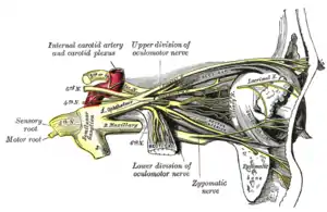
 Superior view of the nerves of the orbit. The lacrimal nerve is seen branching from the ophthalmic nerve.
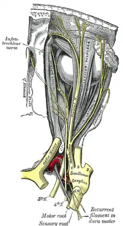
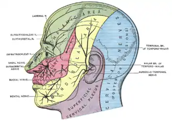 Sensory innervation to the skin of the head and neck. The cutaneous distribution of the lacrimal nerve can be seen above the eye in the green area.
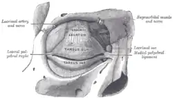 Anterior view of the orbit and tarsal plates. The lacrimal nerve can be seen exiting the orbit superolaterally after it supplies the lacrimal gland.
 Superior view of a dissection of the left orbit. The lacrimal nerve is visible innervating the lacrimal gland.
References
- Standring, Susan (2020). Gray's Anatomy: The Anatomical Basis of Clinical Practice (42th ed.). New York. p. 631. ISBN 978-0-7020-7707-4. OCLC 1201341621.
{{cite book}}: CS1 maint: location missing publisher (link) - Sinnatamby, Chummy S. (2011). Last's Anatomy (12th ed.). Elsevier Australia. ISBN 978-0-7295-3752-0.
- Standring, Susan (2020). Gray's Anatomy: The Anatomical Basis of Clinical Practice (42nd ed.). New York. p. 782. ISBN 978-0-7020-7707-4. OCLC 1201341621.
{{cite book}}: CS1 maint: location missing publisher (link)