YjeF N terminal protein domain
In molecular biology, the YjeF N terminal is a protein domain found in the N-terminal of the protein, EDC3. The YjeF N-terminal domains occur either as single proteins or fusions with other domains and are commonly associated with enzymes. They help assemble the processing body (P-body) in preparation for mRNAdecay. Structural homology indicated it may have some similarity to the enzyme family, hydrolase.
| YjeF_N | |||||||||
|---|---|---|---|---|---|---|---|---|---|
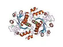 crystal structure of yeast ynu0, ynl200c | |||||||||
| Identifiers | |||||||||
| Symbol | YjeF_N | ||||||||
| Pfam | PF03853 | ||||||||
| InterPro | IPR004443 | ||||||||
| SCOP2 | 1jzt / SCOPe / SUPFAM | ||||||||
| |||||||||
Function
At the cellular level, the YjeF-N terminal domain is vital to the assembly of the processing body (P-body). This aids mRNA decay and is thought to bring together different complexes to aggregate mRNPs.[1] At the organism level, in bacteria and archaea, YjeF N-terminal domains are often fused to a YjeF C-terminal domain with high structural homology to the members of a ribokinase-like superfamily or belong to operons that encode enzymes of diverse functions. Examples of such include:
- pyridoxal phosphate biosynthetic protein PdxJ;
- phosphopanteine-protein transferase;
- ATP/GTP hydrolase;
- and pyruvate-formate lyase 1-activating enzyme.
In plants, the YjeF N-terminal domain is fused to a C-terminal putative pyridoxamine 5'-phosphate oxidase. In eukaryotes, proteins that consist of (Sm)-FDF-YjeF N-terminal domains may be involved in RNA processing.[2][3]
Structure
The YjeF N-terminal domains represent a novel version of the Rossmann fold, one of the most common protein folds in nature. The YjeF N-terminal domain is a three-layer alpha-beta-alpha sandwich with a central beta-sheet surrounded by alpha helices. The conservation of the acidic residues in the predicted active site of the YjeF N-terminal domains shows some similarities to the amino acids found in the active sites of diverse hydrolases.[2][3]
References
- Ling SH, Decker CJ, Walsh MA, She M, Parker R, Song H (2008). "Crystal structure of human Edc3 and its functional implications". Mol Cell Biol. 28 (19): 5965–76. doi:10.1128/MCB.00761-08. PMC 2547010. PMID 18678652.
- Anantharaman V, Aravind L (July 2004). "Novel conserved domains in proteins with predicted roles in eukaryotic cell-cycle regulation, decapping and RNA stability". BMC Genomics. 5 (1): 45. doi:10.1186/1471-2164-5-45. PMC 503384. PMID 15257761.
- Jha KN, Shumilin IA, Digilio LC, Chertihin O, Zheng H, Schmitz G, Visconti PE, Flickinger CJ, Minor W, Herr JC (May 2008). "Biochemical and structural characterization of apolipoprotein A-I binding protein, a novel phosphoprotein with a potential role in sperm capacitation". Endocrinology. 149 (5): 2108–20. doi:10.1210/en.2007-0582. PMC 2329272. PMID 18202122.